Brain Development, Drugs and Disease
Brain structure
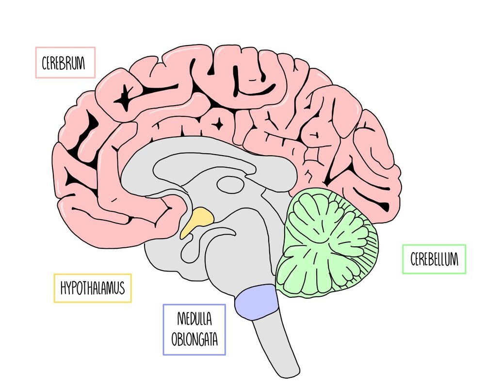
The brain can be divided into specific regions which have specific functions. Most of our brain is made up of the cerebrum, which is found at the top of the brain. It is divided into two cerebral hemispheres joined together by a band of nerve fibres called the corpus callosum. The thin, outer layer of the cerebrum is called the cerebral cortex. It is highly folded, giving it a really large surface area. The cerebrum is involved in ‘higher-brain functions’, such as processing language, vision, thinking and emotions.
Below the cerebrum is a structure called the hypothalamus, which is involved in homeostatic responses such as maintaining body temperature (thermoregulation). It also produces hormones that control the pituitary gland, which is found just beneath the hypothalamus.
The cerebellum is a leaf-shaped structure found towards the back of the brain. It is positioned underneath the cerebrum and is highly folded. It plays an important role in movement and balance. Things like learning to ride a bike or the movement involved in writing will involve a large input from the cerebellum.
Right at the base of the brain and above the spinal cord is a structure called the medulla oblongata. This is involved in unconscious processes, such as the regulation of breathing rate and heart rate.
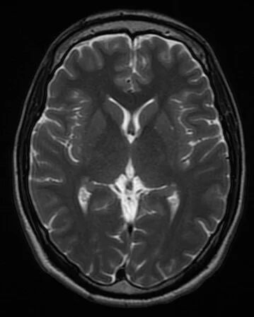
CT scans

CT scan of the brain, taken from the top of the skull to its base. Credit: Department of Radiology, Uppsala University Hospital.
Computed tomography (CT) scans are images of the brain which can be taken to view the structure of the brain and to assess whether brain damage has occurred.
Use X-ray radiation to produce a cross-section image of the brain.
Denser brain structures absorb more X-ray radiation and show up as darker regions on the image.
Useful for diagnosing medical conditions because they can indicate areas of disease or damage. For example, bleeding in the brain is visible on CT scans because blood had a different density to brain tissue.
Although you cannot use CT scanners to work out the function of different brain regions directly, you can infer the functions of different brain regions by matching a patient’s symptoms with areas of brain damage. For example, if a CT scan of a person with dementia shows damage to the cerebrum, this indicates that the cerebrum plays a role in the consolidation of memories.
MRI scans
MRI scan of a healthy brain. Credit: Novaksean, CC BY-SA 4.0 https://creativecommons.org/licenses/by-sa/4.0, via Wikimedia Commons
Magnetic resonance imaging (MRI) scanners use radio waves in the presence of a strong magnetic field to produce cross-sectional images of the brain.
Higher quality and have a higher resolution than CT scans.
Also used in medical diagnosis because they can be used to visualise damaged or diseased parts of the brain. For example, MRI scans can be used to indicate the presence of a brain tumour, which shows up as a lighter colour on an MRI scan.
Doctors can determine the size and location of the tumour and decide on the best treatment option to take. Just like CT scanning, the function of brain regions can be inferred by matching up a patient’s symptoms with areas of brain damage.
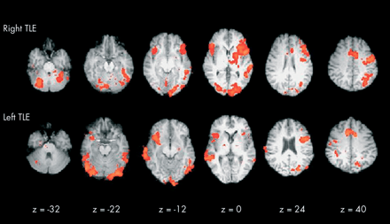
Areas in red show the activation of brain regions when an individual with temporal lobe epilepsy (TLE) carries out a verbal fluency task. Credit: Matthews PM, Jezzard P. Functional magnetic resonance imaging. Journal of Neurology, Neurosurgery & Psychiatry 2004;75:6-12.
fMRI scans
Function magnetic resonance imaging (fMRI) scanners show which areas of the brain are active by detecting changes in blood flow.
Brain regions which are stimulated will be respiring more, so more blood will be flowing to these regions (to deliver more glucose and oxygen).
Oxygenated blood produces a stronger signal in a magnetic field compared to deoxygenated blood, so these areas show up on the fMRI scan.
fMRI scans are similar to MRI scans but they can also be used to research the function of different brain structures. For example, a person inside the scanner may be asked to look at images of different faces. The areas of the brain which light up on the fMRI scan will indicate the brain regions which are involved in facial recognition. fMRI scans are also used in medical diagnosis since they show damaged and diseased parts of the brain.
PET scans
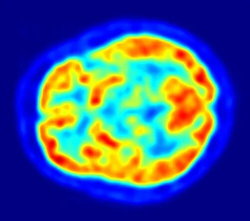
PET scan of a healthy brain. Credit: By Jens Maus (http://jens-maus.de/) - Own work, Public Domain
Like fMRI, positron emission tomography (PET) scans also show which regions of the brain are activated at any given time, but they use radioactive tracers such as radioactively labelled glucose.
The radioactively labelled glucose will accumulate in parts of the brain which are respiring more and will produce a stronger signal on the PET scan.
Just like MRI scans, PET scans have a high resolution and high quality.
They can be used to determine the function of different brain regions and can also identify areas of brain damage or disease.
Habituation
Habituation is a kind of learned response where organisms learn to ignore unimportant stimuli (i.e. they know that they are not dangerous or do not offer any reward) after repeated exposure. It’s what happens if you live close to a hospital and start to blank out the sound of sirens, for example. It’s important because it means that organisms don’t waste time and energy by responding to stimuli that do not pose a danger to them.
When habituation occurs, the action potentials that result from the stimulus dampen down over time. The repeated exposure to the stimulus decreases the amount of calcium ions which enter the presynaptic neurone, which means that fewer vesicles containing neurotransmitters release their contents into the synaptic cleft. This means that there are less neurotransmitters to bind to receptors on the postsynaptic neurone, so less sodium ions channels open in the postsynaptic neurone. Less depolarisation of the membrane occurs, which may not reach the threshold potential. This means that fewer action potentials will reach the effector (the muscle or the gland) which carries out the response.
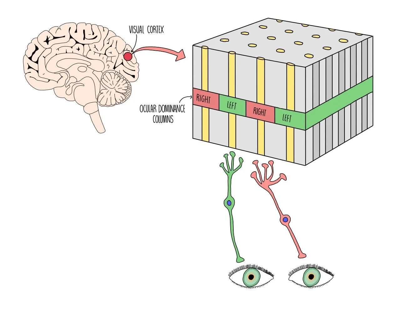
Visual cortex
The visual cortex is a region at the back of our brains and forms part of the cerebral cortex. It helps us to process visual information. Neurones in the visual cortex receive information from either our right or left eye and are clustered together in structures called ocular dominance columns. Right ocular dominance columns receive information from our right eye while left ocular dominance columns receive information from our left eye. The ocular dominance columns are arranged within the visual cortex in a repeating alternating pattern (i.e. right, left, right, left, and so on).
Development of the visual cortex
When we are born, our brains contain a load of neurones which begin to form connections (synapses) or are removed, making the brain more organised and allowing our visual system to develop. This all happens very early in life and relies on both of our eyes receiving visual input. The period of early life when our brains are developing is called the critical period. During the critical period, synapses that receive visual stimulation and pass on action potentials into the visual cortex are retained and strengthened. Synapses that do not receive visual stimulation, so the neurones between them are not firing, are removed. This means that if visual stimulation does not occur during the critical period (i.e. if a baby is born with cataracts which obscure vision or if they are born in a cave) then their visual cortex will not develop properly because many of the synapses will have been destroyed. Evidence for a ‘critical period’ comes from some ethically-dubious experiments on kittens (see below).
Hubel and Wiesel’s experiment
David Hubel and Torsten Wiesel were two scientists who studied the electrical activity of neurones in the visual cortex of different animals. They were the first to discover the presence of ocular dominance columns and they determined that both right and left ocular dominance columns exist which are stimulated by visual input from the right and left eyes respectively.
Hubel and Wiesel carried out an experiment on kittens and adults cats in 1963 which proved that the visual cortex develops during a critical period early in life. First, they prevented visual stimulation in one eye by sewing up one eye of each kitten. Several months later, they unstitched the eye and found that the kittens had gone blind in one eye. Looking at the brains of the kittens under the microscope, they found that the ocular dominance columns for the stitched up eye had shrunk and that the ocular dominance columns for the open eye had expanded, suggesting that the columns for the open eye had taken over the columns that were not being stimulated (i.e. the neurones in the visual cortex had switched dominance). When they repeated the experiments in adult cats, they found that the cats retained their normal vision after having their eye stitched closed for several months and their ocular dominance columns remained unchanged. They repeated the experiment on young and adult monkeys and achieved the same results. Their results showed that the visual cortex only develops normally if both eyes receive visual stimulation in early life.
Since the visual cortex in cats and humans is similar (they both contain ocular dominance columns), Hubel and Wiesel’s results can be applied to humans. This means that humans need to receive visual stimulation from both eyes during the early periods of life for their visual cortex to develop normally. For example, some babies are born with cataracts which make the lens of the eye go cloudy and obscures vision. Unless the cataracts are removed, the baby’s visual cortex will not develop properly because they are not receiving sufficient visual stimulation during early life. However, if adults develop cataracts it will not affect their visual system because it has already developed.
Ethical issues with animal studies
Animals are commonly used in medical research as they enable us to test theories and ensure that drugs are safe to use, meaning that many human lives are saved as a result. However, their use is controversial and there are a number of arguments against their use. Some of the arguments for and against the use of animals in medical research are summarised below:
Arguments for the use of animal research:
It allows us to test the effects on a whole organism. An alternative to using animals is cell culture but this has the disadvantage that the cells are not part of a whole system - other body tissues may play a role in physiological responses.
It allows us to carry out research which would be unethical on humans and helps us to develop effective drugs which save countless human lives.
Animal experiments are only carried out when necessary and scientists have to follow strict guidelines and procedures e.g. animals need to be well looked after and euthanized humanely.
The similarities between animals and humans mean that we can apply the results to humans, making them an invaluable research tool.
Arguments against the use of animal research:
All organisms have the right to not suffer and it is unethical to cause pain or stress to any living creature.
There are alternatives to using animals - i.e. cell culture or computer models.
Animals and humans are not the same and there is the possibility (indeed, this quite often happens) that drugs which are safe in animals have a different response and side effects in humans.
Studying brain development
The way that our brains develop is a result of both our genes (nature) and our environment (nurture). Some scientists disagree about the extent that nature or nurture influence the development of the brain - this is known as the nature-nurture debate. There are five different types of study which scientists can use to work out the role that nature and nurture play in brain development:
Twin studies - identical twins that have been raised in different environments (i.e. adopted by separate families) can show the influence of nature vs nurture. Because identical twins have the same genes, any similarities will be due to nature and any differences due to nurture. For example, identical twins that have been separated have very similar IQ scores, suggesting that nature has a large influence in intelligence. Scientists can also compare identical twins (who haven’t been separated - so same genes, same environment) with non-identical twins (different genes, same environment). Any differences between the identical and non-identical twins will be due to nature. For example, stuttering in both twins is more identical among identical compared to non-identical twins, suggesting that speech/stuttering is largely influenced by nature.
Cross-cultural studies - scientists can look at data from large groups of children who have been brought up in different cultures. Any differences between them will largely be due to nurture rather than nature. For example, mapping and spatial abilities of young children is similar across cultures, suggesting that this ability is largely due to nature.
Animal experiments - animals of the same species have similar genes, so brain development can be compared between animals of the same species, with any differences being due to nurture than nature. For example, rats raised in a stimulating environment have larger brains and are better at problem-solving than rats raised in a dull environment, suggesting that nurture plans a role in brain size and ability to solve problems. Scientists can also genetically engineer mice that are missing certain genes to determine the effect of those genes on brain development. If scientists knock out a gene called Lgl1, the brain expanded and is filled with fluid. This suggests that nature plays a large role in normal brain development.
Newborn studies - apart from the womb, newborn babies have had little exposure to the environment so any abilities that they are born with (i.e. the ability to cry) is likely to be innate and due to nature rather than nurture. Any abilities which take more time to develop (such as language) are more likely to be due to nurture.
Brain damage - scientists can compare children with and without brain damage to determine which abilities are due to nurture. If a child with brain damage still develops a certain ability, then that skill will be a result of nurture. For example, children with brain damage to the region of the brain associated with language initially show impairments in speaking but by the age of five there is no difference between these children and healthy children. This suggests that speaking and processing language has a mostly environmental component (nurture).
Neurotransmitter deficiency diseases
Some diseases are known to be causes by a lack of particular neurotransmitters. Two examples of this kind of disease is Parkinson’s, which is caused by a lack of dopamine in the brain, and depression, which is caused by a lack of serotonin.
Parkinson’s disease is a movement disorder resulting from the destruction of neurones in regions of the brain which control movement. Neurones in this region of the brain produce dopamine, so neuronal death in this part of the brain results in a lack of dopamine. This means that when an action potential arrives at a neurone, the presynaptic neurone releases less dopamine into the synaptic cleft. Less dopamine receptors on the postsynaptic neurone are activated so less sodium ion channels in the postsynaptic membrane open. This makes it less likely for the threshold potential in the postsynaptic neurone to be reached, so a nerve impulse is less likely to fire. The reduction in neuronal transmission in parts of the brain associated in movement results in symptoms such as tremors and poor motor coordination. Parkinson’s disease is treated with drugs which increase levels of dopamine in the brain.
L-dopa is the main drug treatment for Parkinson’s disease. It has a similar structure to dopamine and is able to cross the blood-brain barrier and pass into the brain, where it is converted into dopamine by the enzyme dopa-decarboxylase. Dopamine cannot be given directly to patients since it cannot pass through the blood-brain barrier). L-dopa therefore increases dopamine levels in the brain, resulting in more nerve impulses along neurones in brain regions which are involved in movement.
Depression is a mood disorder which is thought to be caused, in part, by a lack of serotonin in the brain. Antidepressants have been developed to increase the levels of brain serotonin. A class of antidepressants known as selective serotonin reuptake inhibitors (SSRIs) work by binding to serotonin reuptake proteins within synapses, blocking the proteins and preventing them from reabsorbing serotonin.
Another substance which increases the brain’s serotonin levels is the party drug MDMA (ecstasy). MDMA prevents the neurone’s ability to reabsorb serotonin from synapses by binding to and blocking reuptake proteins on the presynaptic membrane. It also stimulates presynaptic neurones to release more serotonin. This increases serotonin levels in the brain and increases the frequency of nerve impulses along neurones in brain regions which are involved in mood.
Using genome sequencing to produce drugs
The Human Genome Project (HGP) was an ambitious target, set in 1990, to sequence the entire DNA found in humans. It took 13 years and has been used as an invaluable tool in medicine. Scientists are able to use the database to identify genes and proteins which are implicated in disease. This information can then be used to create new drugs to target those proteins. Genome sequencing has also identified tiny genetic variations between people where just one nucleotide differs - the fancy term for this is single nucleotide polymorphisms (SNPs). Some of these genetic variations are known to make some drugs less effective. This has resulted in a new area of healthcare called personalised medicine where doctors can prescribe a unique treatment plan depending on the genetic variations found in each individual patient.
However, there are social, moral and ethical issues for personalised medicine. There is the danger that a person’s genetic information may be passed onto third parties and used against them (e.g. if an insurance company knows that you have a mutation that is associated with breast cancer, or a SNP which means you may not respond well to a particular treatment, they may choose to charge more for a health insurance plan). There is also the concern that tailored medicine will increase research costs, making drugs more expensive and only affordable for more wealthy patients. In addition, more expensive drugs may be denied to certain patients on the grounds that they possess an SNP which indicates that they may not respond well to a treatment. Finally, if a patient knows that they have an SNP which means they may not respond well to treatment, it might have a negative psychological effect which means that the drugs responds worse than if they hadn’t been told about their genetic variation at all (kind of like a reserve-placebo effect).
Producing drugs using genetically modified organisms (GMOs)
Genetically modified organisms (GMOs) are organisms which have had their DNA changed so that they synthesise proteins which can be used as drugs. Restriction enzymes are used to cut DNA at specific sites to extract a gene of interest. The same restriction enzymes are used to cut plasmid DNA, creating complementary sticky ends. DNA ligase joins the two pieces of DNA together so that the gene of interest is now contained within a plasmid. This is called recombinant DNA. This is either placed in a virus, which will infect organisms with the recombinant DNA, or the plasmid will be taken up by bacteria. Plasmids and viruses which carry the DNA molecule are called vectors.
This process is used for the production of insulin.
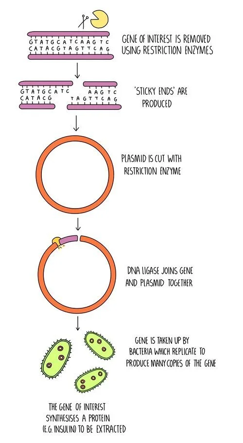
The insulin gene is removed from human DNA using restriction enzymes.
A plasmid is also cut with restriction enzymes.
DNA ligase joins the complementary sticky ends to form the recombinant DNA.
The recombinant plasmid is taken up by bacteria.
The transgenic bacteria are grown in large fermenters to produce large amounts of insulin, which can then be extracted.
Bacteria aren’t the only organisms which can be genetically modified to produce drugs - plants and animals can be used too. For genetically modifying plants, a GM bacterium is first made using the process outlined above. The bacterium then acts as a vector, infecting a plant cell and inserting its DNA into the genome of the plant cell. The plant cell grows into an adult plant and all of its cells will contain the drug-producing gene. The plant cells will synthesise the protein which can either be extracted or the drug can be delivered to the patient by eating the plant. Cholera vaccines have been made using this method.
Genetically-modified animals are produced by injecting the gene for the protein (which will act as the drug) into the nucleus of a fertilised animal egg cell. This is then implanted into an adult animal and as the animal develops, every cell will contain the drug-producing gene. The protein produced from the gene is normally purified from the milk of the animal. This method has been used in goats to produce the drug antithrombin for treating people with defective blood clotting.
Issues with using GMOs in medicine and food production
GMOs are not just used for producing drugs, but also for improving agriculture. For example, genes can be added to crop plants to make them resistant to disease or more nutritious. However, the use of GMOs is controversial and there are some arguments for and against their use in medicine and agriculture:
Arguments for the use of GMOs:
Genetic modification can increase crop yield and make food more nutritious - this means that farmers can make more money and people will be healthier.
Genes for pest-resistance can be introduced into crops. This means that farmers save money on pesticides and the environmental problems associated with pesticide use are reduced.
Human proteins produced using GMOs do not result in allergic reactions - for example, insulin as a treatment for diabetes used to be extracted from the blood of pigs and cows. Nowadays human insulin is produced using GM bacteria which doesn’t produce an allergic reaction in patients and is more efficient.
Vaccines produced using genetically-modified plants do not need to be kept cold to stay effective - this is an advantage when vaccines are needed in remote regions where refrigeration is not possible.
Enzymes can be produced using GMOs and are often used in industrial processes e.g. in the manufacture of washing detergents and the textile industry. The use of GMOs makes these processes cheaper.
The process is cheap because once one genetically modified organism is made, lots more can be made through breeding the original GM organism. This makes drugs cheaper.
Arguments against the use of GMOs:
GMOs are often patented and the seeds are expensive to buy - this puts farmers in developing countries at a disadvantage.
If a GM plant cross-pollinated with another species, it could introduce the genes into other plants with unintended consequences. For example, cross-breeding a herbicide-resistant crop plant with wild plant species could create ‘superweeds’ which are resistant to herbicides. These could out-compete other plants with potential effects on the whole food chain.
There are ethical issues against the use of GMOs - should humans manipulate animals for our own benefit?
Religious reasons - do humans have the right to ‘play God’ and create new life?
Some people worry out the long-term impact of using GMO - there may be unforeseen consequences which are impossible to predict.