Cell Structure and Mitosis
Ultrastructure of eukaryotic cells
All organisms are divided into two different domains: eukaryotes and prokaryotes. Eukaryotes include any organism whose cells contain a nucleus, while prokaryotes lack a nucleus and any other membrane-bound organelles.
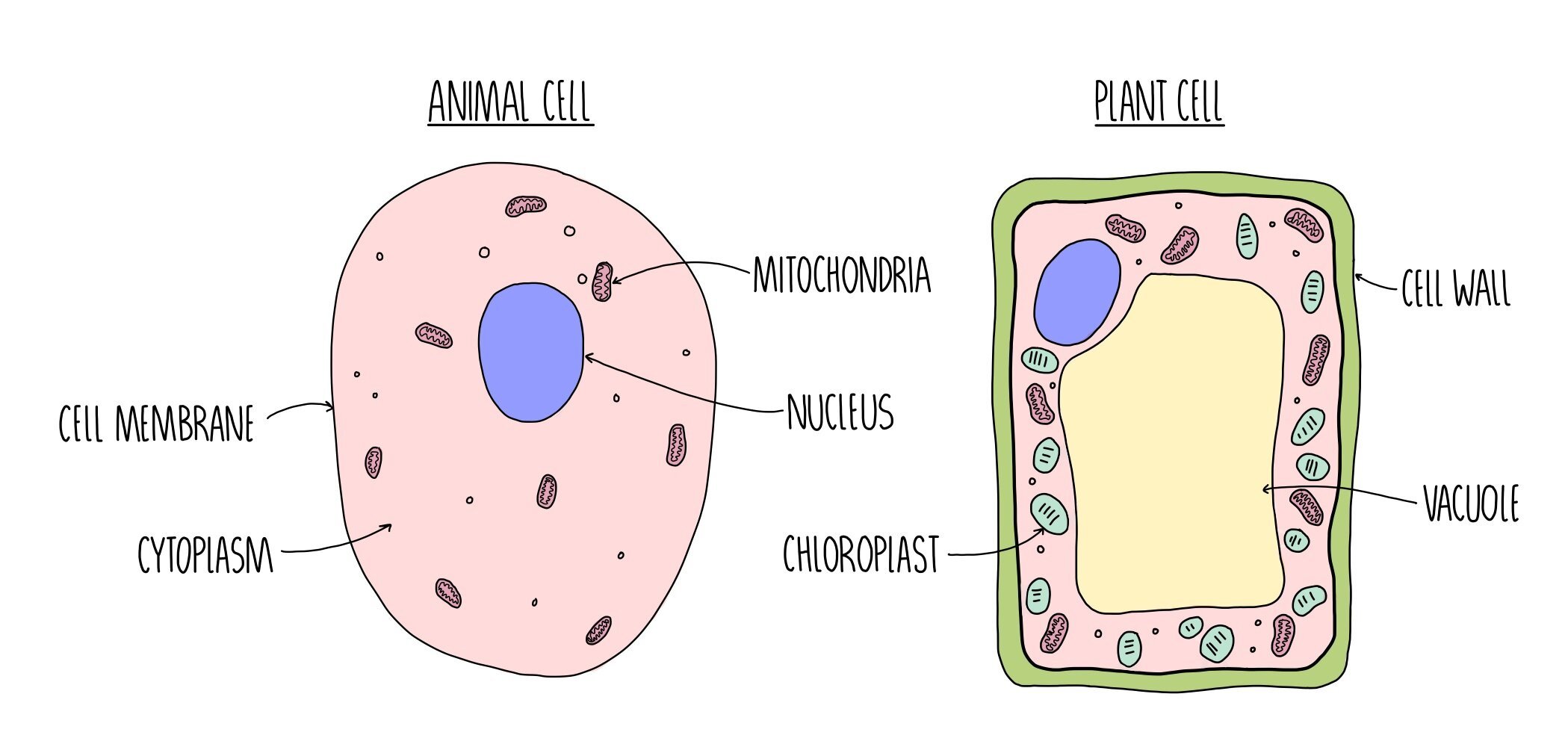
Eukaryotic cells, such as the cells of animals, plants and fungi may contain the following organelles:

CHLOROPLAST
MITOCHONDRIA

GOLGI APPARATUS
Nucleus - contains DNA which controls the activities of the cell by containing the base sequences (the ‘instructions’ needed to make proteins). The DNA is associated with histone proteins and referred to as chromatin which is wound into structures called chromosomes.
Nucleolus - this is a region within the nucleus where ribosomes are made.
Nuclear envelope - a double membrane which surrounds the nucleus. It contains pores which allows small molecules (like single stranded RNA) to pass into the cytoplasm but keeps hefty chromosomes safely inside its walls.
Rough endoplasmic reticulum (RER) - the RER is an extension of the nuclear envelope and is coated with ribosomes. It facilitates protein synthesis by providing a large surface area for ribosomes. It then transports the newly synthesised proteins to the Golgi apparatus for modification.
Smooth endoplasmic reticulum (SER) - synthesises lipids including cholesterol and steroid hormones (such as oestrogen).
Golgi apparatus - made up of a group of fluid-filled membrane-bound flattened sacs surrounded by vesicles. It receives proteins from the RER and lipids from the SER. It modifies the proteins and lipids and repackages them into vesicles. The Golgi apparatus is also the site of lysosome synthesis.
Ribosomes - ribosomes are responsible for the translation of RNA into protein (protein synthesis). They either float freely in the cytoplasm or are stuck onto the rough endoplasmic reticulum.
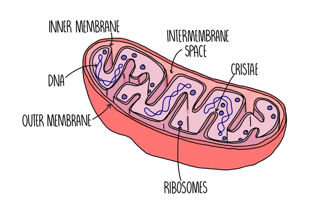
Mitochondria - site of ATP production during aerobic respiration. It is self-replicating so can become numerous in cells with high energy requirements. It contains a double membrane with folds called cristae, which provides a large surface area for respiration.
Lysosomes - phospholipid rings which contain digestive enzymes separate from the rest of the cytoplasm. Lysosomes engulf and destroy old organelles or foreign material.
Chloroplasts - the site of photosynthesis. It is enclosed by a double membrane and has internal thylakoid membranes arranged in stacks to form grana linked by lamellae. These structures are found only in plants and certain types of photosynthesising bacteria or protoctists.
Plasma membrane - consists of a phospholipid bilayer with additional proteins to serve as carriers. It also contains cholesterol to regulate membrane fluidity. The plasma membrane contains the cell contents and holds the cell together, whilst controlling the movement of substances into and out of the cell.
Centrioles - these are bundles of microtubules which form spindle fibres during mitosis in order to pull sister chromatids apart. They are also important for the formation of cilia and flagella. They are not found in plant and bacterial cells.
Cell wall - a rigid structure made of cellulose (in plants), chitin (in fungi) and murein (in prokaryotes) which provide support to the cell.
Flagella - a tail-like structure which are made up of bundles of microtubules. The microtubules contract to make the flagellum move and propel the cell forward. Cells with a flagellum include sperm cells, which use it to swim up the fallopian tubes to fertilise the egg cell.
Cilia - finger-like projections found on the surface of some cells. These also contain bundles of microtubules which contract to make the cilia move. Cilia are found on epithelial cells lining the trachea and move to sweep mucus up the windpipe.
Vacuole - the vacuole is an organelle which stores cell sap and may also store nutrients and proteins. It helps to keep plant cells turgid. Some vacuoles can perform a similar function to lysosomes and digest large molecules.
Protein production
Translation of mRNA into a polypeptide chain takes place on ribosomes which are either floating alone in the cytoplasm or attached to the rough ER.
The long polypeptide chain is folded at the rough ER and transported to the Golgi apparatus inside vesicles.
At the Golgi, they are modified and processed by various enzymes. The protein may have a carbohydrate chain stuck onto its surface, or the addition of a sulfate or phosphate group.
It is packaged inside another vesicle which travels to the part of the cell where the protein is needed. If the protein is a carrier protein, the vesicle will deliver the protein to the plasma membrane where it will be incorporated.
Comparing eukaryotic and prokaryotic cells
Prokaryotes and eukaryotes share some of the same organelles (cytoplasm, cell membrane, ribosomes) but there are some important differences:
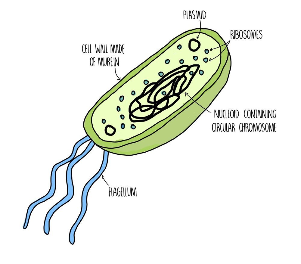
Prokaryotes have no membrane-bound organelles (so no mitochondria, Golgi, endoplasmic reticulum, nucleus etc.). Their DNA floats freely in the cytoplasm.
Their DNA consists of a single circular chromosome whereas DNA in eukaryotes is linear and wrapped around chromosomes.
Prokaryotes have extra bits of DNA in the form of small circular plasmids.
Prokaryotes have smaller ribosomes (70S) compared to eukaryotic ribosomes (80S).
Eukaryotes like plants and fungi have cell walls made of cellulose and chitin. Bacterial cell walls are made of murein (a type of glycoprotein).
Prokaryotic cells are much smaller than eukaryotic cells.
Both prokaryotes and eukaryotes can have flagella but those found in prokaryotes are made of a protein called flagellin whereas in eukaryotes they are formed from microtubules.
Prokaryotes have some organelles that are absent from eukaryotic cells. These include:
Pili - pili are hair-like structures which stick out from the plasma membrane. They are used to communicate with other cells (including the transfer of plasmids between bacteria).
Mesosomes - the mesosome is a folded portion of the inner membrane. While some scientists believe that it plays a role in chemical reactions, such as respiration, other scientists doubt whether it even exists and think that it may just be an artefact produced during the preparation of bacterial samples for microscopy.
Plasmids - plasmids are small, circular rings of DNA which are separate from the main chromosome. They house genes which are not crucial for survival but might prove useful - such as antibiotic-resistance genes, for example. Plasmids can replicate independently from the main chromosomal DNA.
Slime capsule - in addition to a cell wall, some bacteria also have a capsule which is made of slime. The main function of the capsule is to protect the bacterium against an immune system attack.
Microscopy
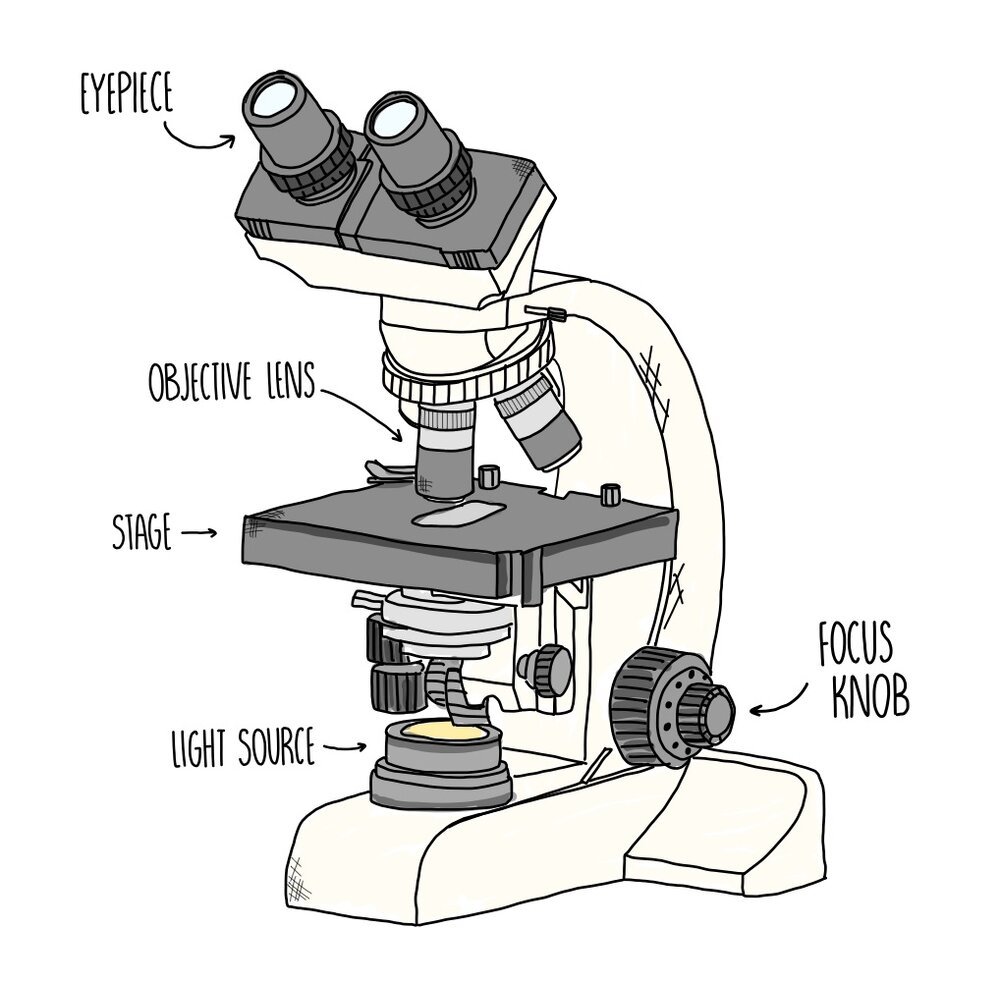
The light microscope uses light to magnify objects up to 1,500x their actual size. They have a resolution of approximately 0.2 μm which isn’t large enough to visualise any of the smaller organelles, such as ribosomes and lysosomes. They are more commonly used for visualising whole cells or tissues. An advantage of light microscopy is that it can visualise living cells so we can watch behaviours such as cell division in real time.
The transmission electron microscope (TEM) is more powerful than a light microscope and has a high enough resolution (around 0.0002 μm) to visualise individual organelles. A TEM uses electromagnets to focus a beam of electrons at a sample. Electrons have a much shorter wavelength compared to visible light which means higher-resolution, detailed images can be produced. A disadvantage of TEM is that the sample needs to be fixed and placed in a vacuum, which means that live cells cannot be used.
The scanning electron microscope (SEM) has a lower resolution (around 0.002 μm) than the TEM but they can produce 3D imagesof cells and organelles. They emit a beam of electrons towards a sample, knocking electrons off it which are used to build an image. Like TEMs, SEMs cannot be used with live cells. Both types of electron microscope are pretty big and expensive so you’ll only find them in specialised research facilities and hospitals.
| Light microscope | TEM | SEM | |
| Maximum magnification | 1,500 x | 1,000,000 x | 500,000 x |
| Maximum resolution | 0.2 um | 0.0002 um | 0.002 um |
Magnification
Magnification is how enlarged the image is compared to the original object. Resolution is defined as how well a microscope distinguishes between two points that are close together (i.e. how much detail it can make out). Light microscopes have a much lower resolution, so produce less detailed images, compared to electron microscopes.
You can work out the magnification of a specimen viewed under a microscope using the equation:

Let’s say we magnify a 2 μm bacterial cell to form an image which is 16 cm long. The magnification we must have used is:
Convert both into the same units. 16 cm = 160 mm = 160,000 μm
160,000 / 2 = 80,000 x magnification
Calibrating the eyepiece graticule and the stage micrometer
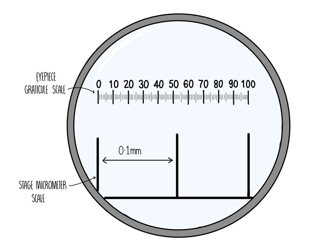
If you wanted to measure the size of your specimen, you’ll first need to align the eyepiece graticule and the stage micrometer which are little rulers which are found on the lens and the stage respectively. To do the calibration you need to carry out the following steps:
Place the stage micrometer on the stage and focus the lens so that you can clearly see the divisions.
Align the eyepiece graticule with the stage micrometer.
Each division of the stage micrometer is 0.1 mm. If the eyepiece graticule spans a total of three divisions, then we know that the total length of the eyepiece graticule is 0.2 mm.
The graticule is divided by a scale from 0 to 100, which means that each individual division is a length of 0.002 mm.
Now we can take away the stage micrometer and add our sample, using the eyepiece graticule to measure its size.
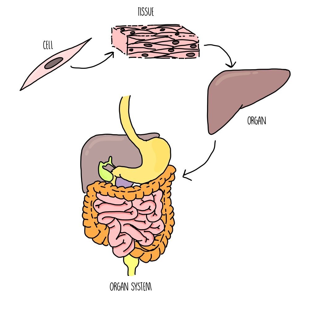
Levels of organisation
The cell is the ‘basic building block of life’ and is the smallest functioning part of an organism. A group of cells working together is called a tissue and a collection of tissues all performing a specific function is called an organ. Multiple organs which are connected together are referred to as an organ system.
Examples of organs include the heart and lungs (in animals) and leaves and roots (in plants).
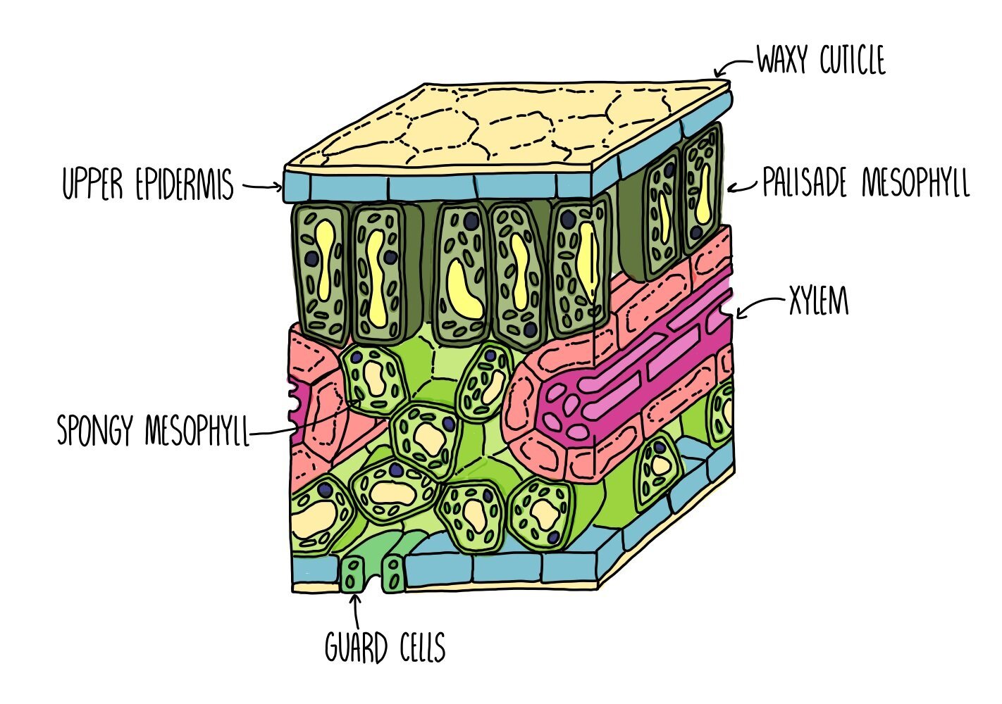
The leaf is an example of a plant organ and is made up of the following tissues:
Upper epidermis – covered in a waterproof waxy cuticle to reduce transpiration.
Palisade mesophyll – tightly packed cells filled with mitochondria, located towards the top of the leaf to absorb as much light as possible. This is where most photosynthesis takes place.
Spongy mesophyll – loosely arranged cells. Air spaces allow circulation of gases for gas exchange.
Phloem – transport of sugars and amino acids (translocation).
Xylem – transport of water and mineral ions (transpiration).
Lower epidermis – contains stomata which open and close to allow gas exchange to take place.
The lungs are an example of an organ found in animals and is made up of the following tissues:
Endothelium – forms the capillary walls which supply alveoli with oxygen and nutrients.
Fibrous connective tissue – helps to push air out of the lungs during exhalation.
Squamous epithelial tissue – makes up the walls of the alveoli.
Examples of organ systems include the respiratory system, circulatory system, reproductive system and digestive system.
Mitosis and the Cell Cycle
Mitosis is a type of cell division where cells produce identical copies of themselves and is used for growth and repair and asexual reproduction. It differs from meiosis, which is the type of cell division used to produce gametes.
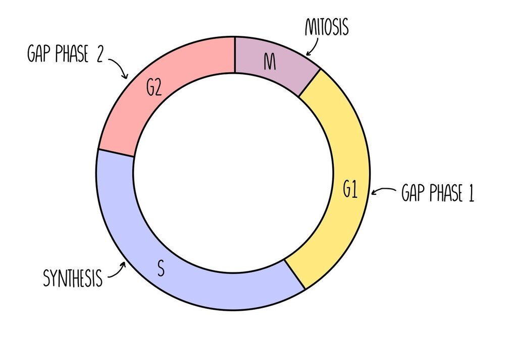
Mitosis occurs as part of the cell cycle which consists of four distinct phases. First, interphase takes place which is made up of three growth phases (called G1 phase, S phase and G2 phase), followed by mitosis.
Gap Phase 1 (G1) - cell grows bigger and replicates its organelles. A high amount of protein synthesis is taking place in order to build new organelles.
Synthesis Phase (S) - the cell replicates its DNA
Gap Phase 2 (G2) - the cell keeps growing until all of the organelles have duplicated.
Once the DNA has replicated, each chromosome now consists of two sister chromatids, connected by a structure called the centromere. The mitochondria produce more ATP which will provide the energy for cell division and the ribosomes will be synthesising a high level of proteins to replicate organelles.

There are two ‘checkpoints’ in the cell cycle - one before S phase and one straight after S phase. During these checkpoints, the cell is checking its DNA for errors. This minimises the chances of duplicating any mutated DNA into the replicated cell.
The Stages of Mitosis
Mitosis can be divided into a series of stages depending on what’s going on with the chromosomes in the cell. You can use the acronym PMAT (pass me another tequila) to help you remember the order.
Prophase - the chromosomes condense (they become shorter and fatter) and the nuclear envelope disintegrates. The centrioles move to opposite poles of the cell and form spindle fibres.
Metaphase - the chromosomes line up along the middle of the cell. They attach to the spindle fibre by their centromere.
Anaphase - the centromere splits and the chromatids are pulled to opposite poles of the cell.
Telophase & cytokinesis - the two groups of chromsomes decondense (they become long and thin) and a nuclear envelope reforms around them, forming two new nuclei. The cytoplasm divides (cytokinesis) and the plasma membrane pinches off to form two new, genetically-identical cells.

Mitotic Index
The mitotic index is a measure of the proportion of cells which are undergoing mitosis. You may be asked to calculate it in the exam. To do this, you need to count the number of cells with visible chromosomes and divide this by the total number of cells.

Investigating mitosis in squashed root tips
You can see mitosis happening in root tip cells by staining the chromosomes and observing under the microscope. We use cells right from the tips of the roots because this is where mitosis is taking place (in the meristem tissue).
Method
Cut a thin section of tissue from the tip of a growing root.
Pipette a set volume of 1M hydrochloric acid into a boiling tube and place in a 60oC water bath.
Place the plant tissue in the boiling tube and leave for five minutes.
Rinse the root tip with cold water and dry using a paper towel.
Cut the root tip so that you have a thin layer of cells (about 2 mm) and spread out onto a microscope slide using a mounted needed.
Add a drop of Toluidine blue O stain to the tissue and place a cover slip on top. Push down on the cover slip to squash the cells and allow light to pass through. Be careful not to push sideways otherwise the chromosomes will become damaged.
Use a light microscope to visualise the cells and identify the stages of mitosis. Any cells with visible chromosomes will be undergoing mitosis (as the chromosomes are condensed).