Eukaryotic and Prokaryotic Cells
Ultrastructure of eukaryotic cells
All organisms are divided into two different domains: eukaryotes and prokaryotes. Eukaryotes include any organism whose cells contain a nucleus, while prokaryotes lack a nucleus and any other membrane-bound organelles.
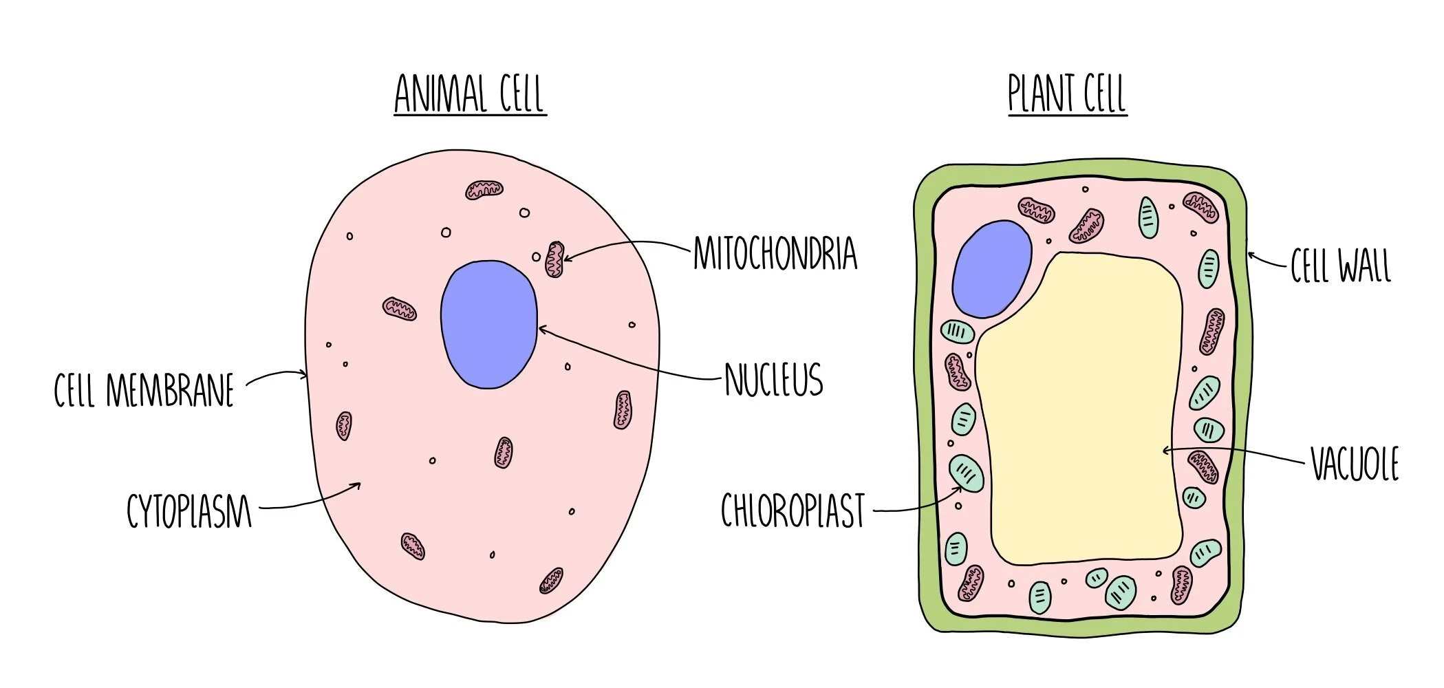
Eukaryotic cells, such as the cells of animals, plants and fungi may contain the following organelles:
Nucleus - contains DNA which controls the activities of the cell by containing the base sequences (the ‘instructions’ needed to make proteins. The DNA is associated with histone proteins and referred to as chromatin which is wound into structures called chromosomes.
Nucleolus - this is a region within the nucleus where ribosomes are made.
Nuclear envelope - a double membrane which surrounds the nucleus. It contains pores which allows small molecules (like single stranded RNA) to pass into the cytoplasm but keeps hefty chromosomes safely inside its walls.
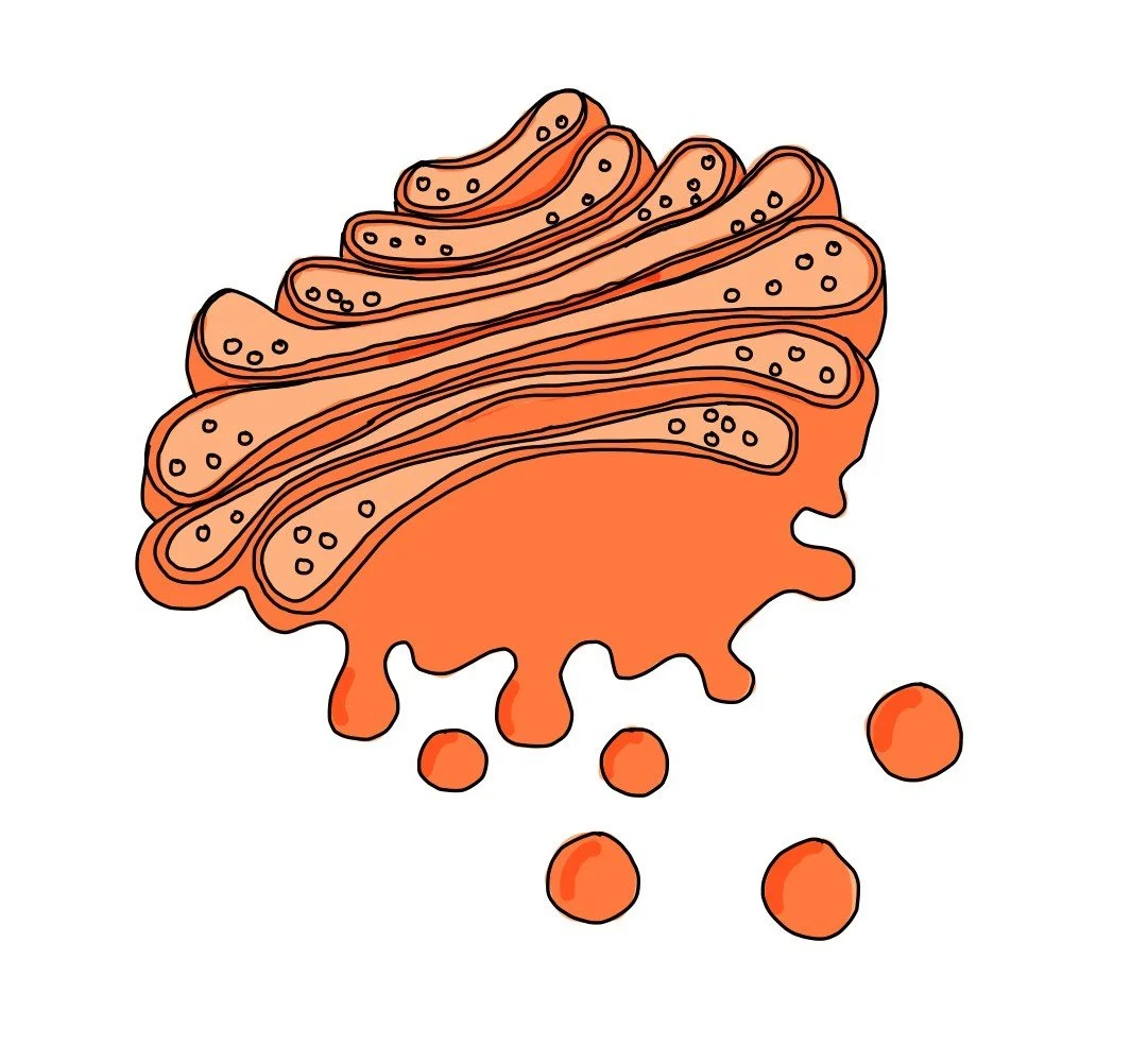
Rough endoplasmic reticulum (RER) - the RER is an extension of the nuclear envelope and is coated with ribosomes. It facilitates protein synthesis by providing a large surface area for ribosomes. It then transports the newly synthesised proteins to the Golgi apparatus for modification.
Smooth endoplasmic reticulum (SER) - synthesises lipids including cholesterol and steroid hormones (such as oestrogen).
Golgi apparatus - made up of a group of fluid-filled membrane-bound flattened sacs surrounded by vesicles. It receives proteins from the RER and lipids from the SER. It modifies the proteins and lipids and repackages them into vesicles. The Golgi apparatus is also the site of lysosome synthesis.
Ribosomes - ribosomes are responsible for the translation of RNA into protein (protein synthesis). They either float freely in the cytoplasm or are stuck onto the rough endoplasmic reticulum.
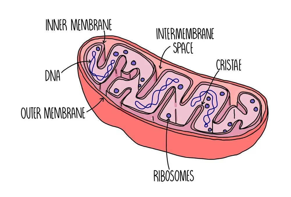
Mitochondria - site of ATP production during aerobic respiration. It is self-replicating so can become numerous in cells with high energy requirements. It contains a double membrane with folds called cristae, which provides a large surface area for respiration.
Lysosomes - phospholipid rings which contain digestive enzymes separate from the rest of the cytoplasm. Lysosomes engulf and destroy old organelles or foreign material.
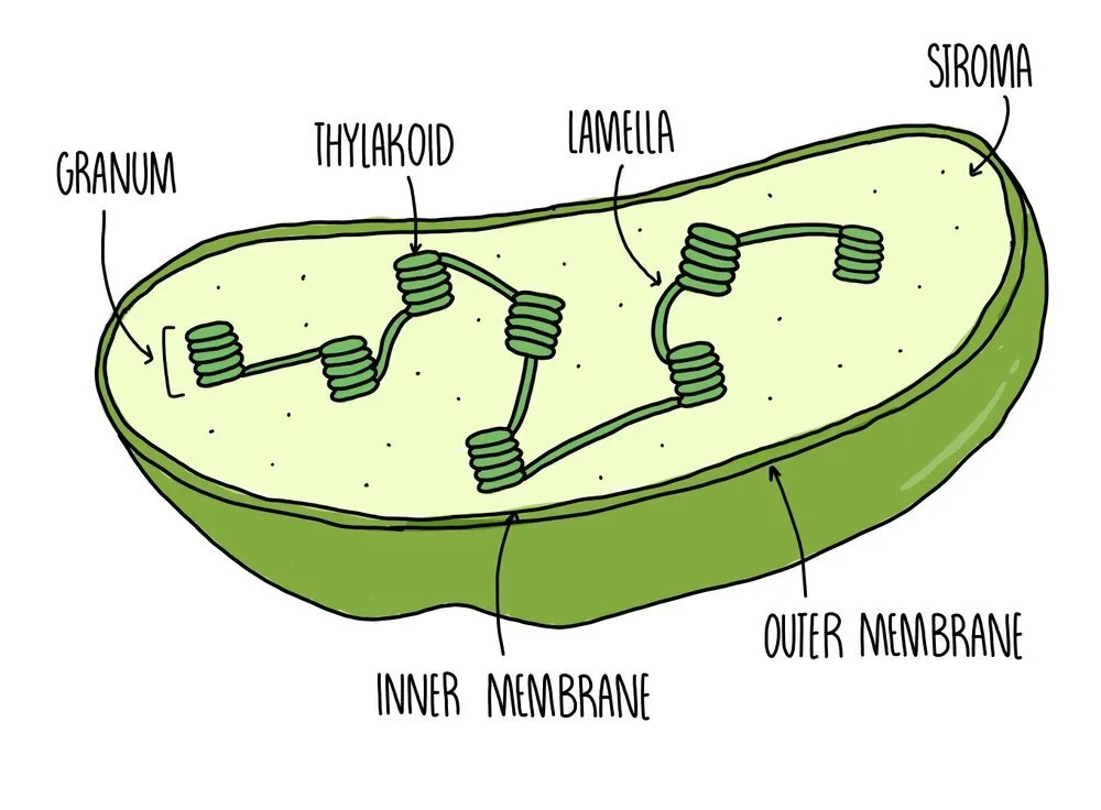
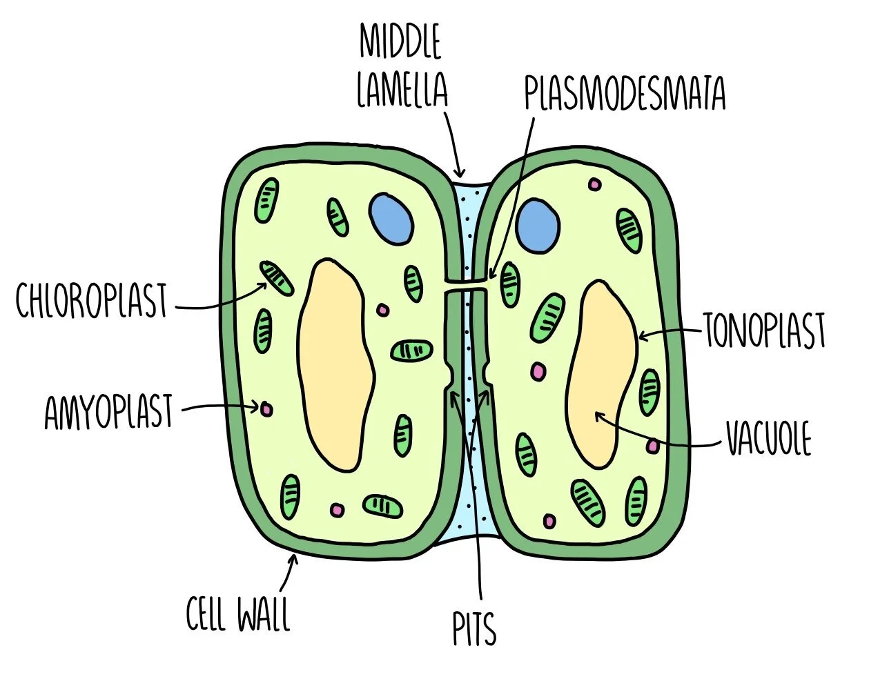
Chloroplasts - the site of photosynthesis. It is enclosed by a double membrane and has internal thylakoid membranes arranged in stacks to form grana linked by lamellae. These structures are found only in plants and certain types of photosynthesising bacteria or protoctists.
Plasma membrane - consists of a phospholipid bilayer with additional proteins to serve as carriers. It also contains cholesterol to regulate membrane fluidity. The plasma membrane contains the cell contents and holds the cell together, whilst controlling the movement of substances into and out of the cell.
Centrioles - these are bundles of microtubules which form spindle fibres during mitosis in order to pull sister chromatids apart. They are also important for the formation of cilia and flagella. They are not found in plant and bacterial cells.
Cell wall - a rigid structure made of cellulose (in plants), chitin (in fungi) and murein (in prokaryotes) which provide support to the cell.
Flagella - a tail-like structure which are made up of bundles of microtubules. The microtubules contract to make the flagellum move and propel the cell forward. Cells with a flagellum include sperm cells, which use it to swim up the fallopian tubes to fertilise the egg cell.
Cilia - finger-like projections found on the surface of some cells. These also contain bundles of microtubules which contract to make the cilia move. Cilia are found on epithelial cells lining the trachea and move to sweep mucus up the windpipe.
Vacuole - the vacuole is an organelle which stores cell sap and may also store nutrients and proteins. It helps to keep plant cells turgid. Some vacuoles can perform a similar function to lysosomes and digest large molecules.
Plasmodesmata - channels (threads of cytoplasm) found between plant cells that enable them to communicate
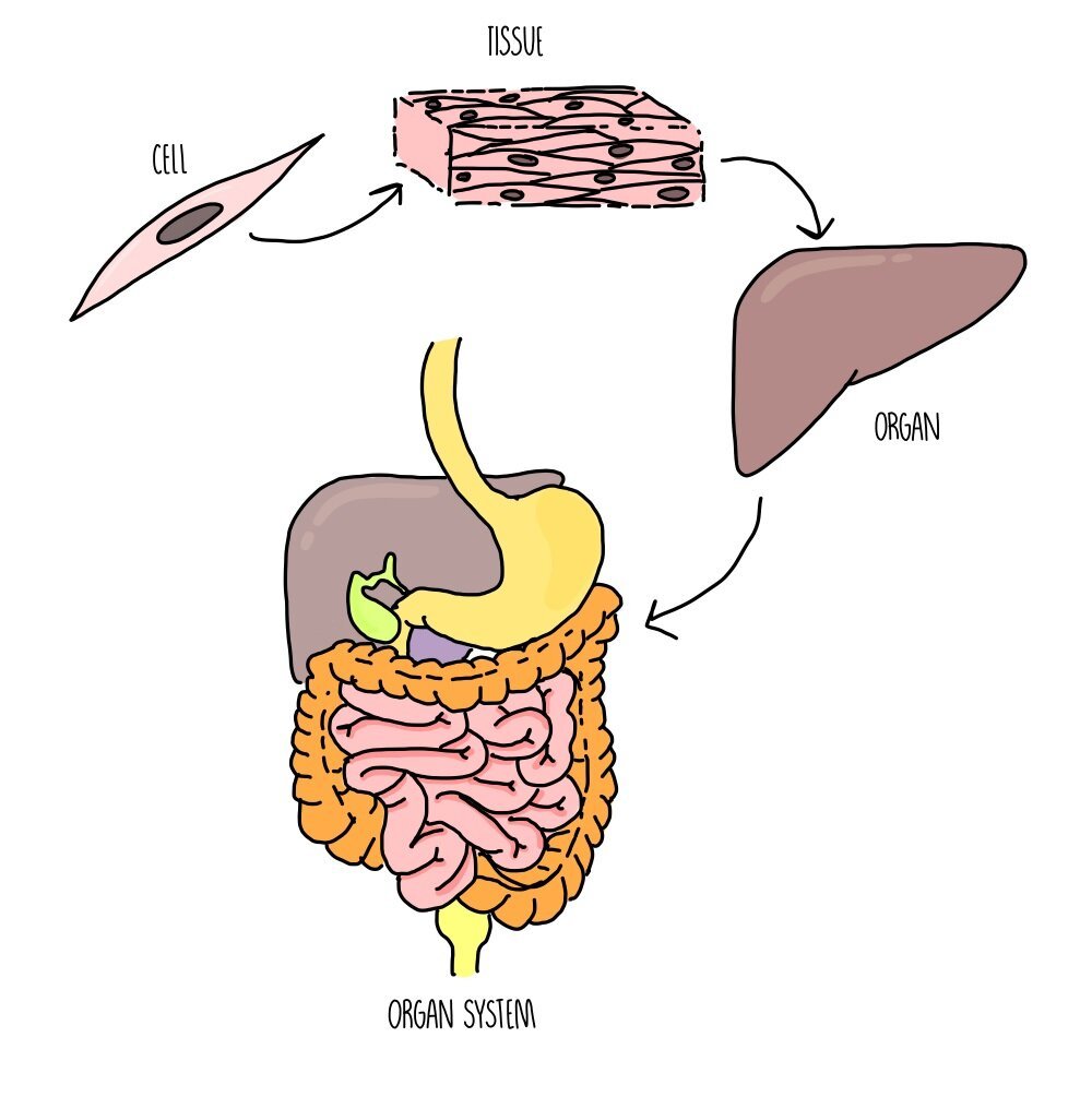
Levels of organisation
The cell is the ‘basic building block of life’ and is the smallest functioning part of an organism. A group of cells working together is called a tissue and a collection of tissues all performing a specific function is called an organ. Multiple organs which are connected together are referred to as an organ system.
The lungs are an example of an organ found in animals and is made up of the following tissues:
Endothelium – forms the capillary walls which supply alveoli with oxygen and nutrients.
Fibrous connective tissue – helps to push air out of the lungs during exhalation.
Squamous epithelial tissue – makes up the walls of the alveoli.
Examples of organ systems include the respiratory system, circulatory system, reproductive system and digestive system.
Comparing eukaryotic and prokaryotic cells
Prokaryotes and eukaryotes share some of the same organelles (cytoplasm, cell membrane, ribosomes) but there are some important differences:
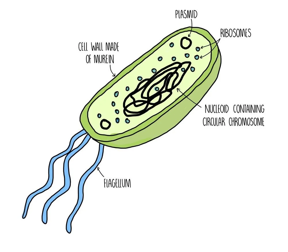
Prokaryotes have no membrane-bound organelles (so no mitochondria, Golgi, endoplasmic reticulum, nucleus etc.). Their DNA floats freely in the cytoplasm.
Their DNA consists of a single circular chromosome whereas DNA in eukaryotes is linear and wrapped around chromosomes.
Prokaryotes have extra bits of DNA in the form of small circular plasmids.
Prokaryotes have smaller ribosomes (70S) compared to eukaryotic ribosomes (80S).
Eukaryotes like plants and fungi have cell walls made of cellulose and chitin. Bacterial cell walls are made of murein (a type of glycoprotein).
Prokaryotic cells are much smaller than eukaryotic cells.
Both prokaryotes and eukaryotes can have flagella but those found in prokaryotes are made of a protein called flagellin whereas in eukaryotes they are formed from microtubules.
Prokaryotes have some organelles that are absent from eukaryotic cells. These include:
Pili - pili are hair-like structures which stick out from the plasma membrane. They are used to communicate with other cells (including the transfer of plasmids between bacteria).
Mesosomes - the mesosome is a folded portion of the inner membrane. While some scientists believe that it plays a role in chemical reactions, such as respiration, other scientists doubt whether it even exists and think that it may just be an artefact produced during the preparation of bacterial samples for microscopy.
Plasmids - plasmids are small, circular rings of DNA which are separate from the main chromosome. They house genes which are not crucial for survival but might prove useful - such as antibiotic-resistance genes, for example. Plasmids can replicate independently from the main chromosomal DNA.
Slime capsule - in addition to a cell wall, some bacteria also have a capsule which is made of slime. The main function of the capsule is to protect the bacterium against an immune system attack.
Bacterial replication
Bacteria replicate by binary fission, which is where the cell replicates its DNA then splits to form two genetically identical daughter cells.
The large loop of chromosomal DNA replicates once. Plasmids also replicate (sometimes more than once). DNA moves to opposite ends of the cell.
The cell grows bigger and cytoplasm divides. New cell wall is synthesised.
The cytoplasm completely splits in two (cytokinesis) and two new cells are formed that are clones of each other (except they may have different numbers of plasmids).
Virus structure
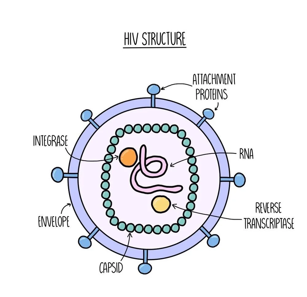
Technically, viruses are not alive because they cannot reproduce on their own (they need to get inside the cells of other organisms to make more copies of themselves).
Contain nucleic acid (either DNA or RNA) and enzymes surrounded by a protein coat called a capsid.
This is enclosed in a viral envelope with attachment proteins sticking out.
Unlike eukaryotic and prokaryotic cells, they do not contain cytoplasm, a cell membrane or ribosomes.
Viral replication
Viruses get inside host cells by binding to receptors on the membranes of host cells using their attachment proteins. This means that viruses can only infect one cell type (which have the complementary shaped receptor on their plasma membrane).
The virus releases its capsid (containing nucleic acids and enzymes) into the cell.
The single strand of nucleic acid is converted into double-stranded DNA and integrated into the host cell’s genome.
The host cell’s ribosomes are used to translate the viral DNA into viral proteins, which are assembled into new viral particles.