Forensics
Microbial decomposition
Microorganism such as bacteria and fungi are able to feed off and decompose dead organic matter - they do this by secreting digestive enzymes onto the organisms.
They digest the tissues of the dead organisms into smaller molecules, such as glucose which is converted into carbon dioxide through respiration.
Methane is also released from microbial decomposition.
They play a very important role in the carbon cycle by unlocking the carbon within glucose and other biological molecules and releasing it into the atmosphere as carbon dioxide gas. The carbon can then be absorbed by plants, converted into glucose by photosynthesis and used to build biological material in living organisms.
Establishing time of death (TOD)
When a dead body turns up under suspicious circumstances, forensic scientists will establish a time of death (TOD). They do this by collecting data on the following factors: extent of body decomposition, the species of insects living on the body, the body’s temperature and how stiff the body is (degree of muscle contraction).
Extent of body decomposition
As soon as a living organism dies, microorganisms will release digestive enzymes which will begin to break down the body tissues into smaller molecules. Depending on the extent of bodily decomposition, a rough TOD can be calculated. However, abiotic conditions such as oxygen availability and environmental temperature will affect how fast microbial decomposition occurs, so this also needs to be factored in. Here’s a rough outline of the stages of decomposition and how these relate to the time of death of a corpse:
From a few hours to a few days - the skin beings to turn green as cells and tissues begin to be broken down
From a few days to a few weeks - methane is given off and the body becomes bloated; skin starts to fall off
After several weeks - tissues liquefy and leak outside of the body
From a few months to a few years - the body tissues have been completely broken down and only a the bones remain
After several decades - the bones disintegrate and nothing remains of the body
Stages of decomposition of a pig carcass.CC BY-SA 3.0, https://commons.wikimedia.org/w/index.php?curid=115198
Forensic entomology
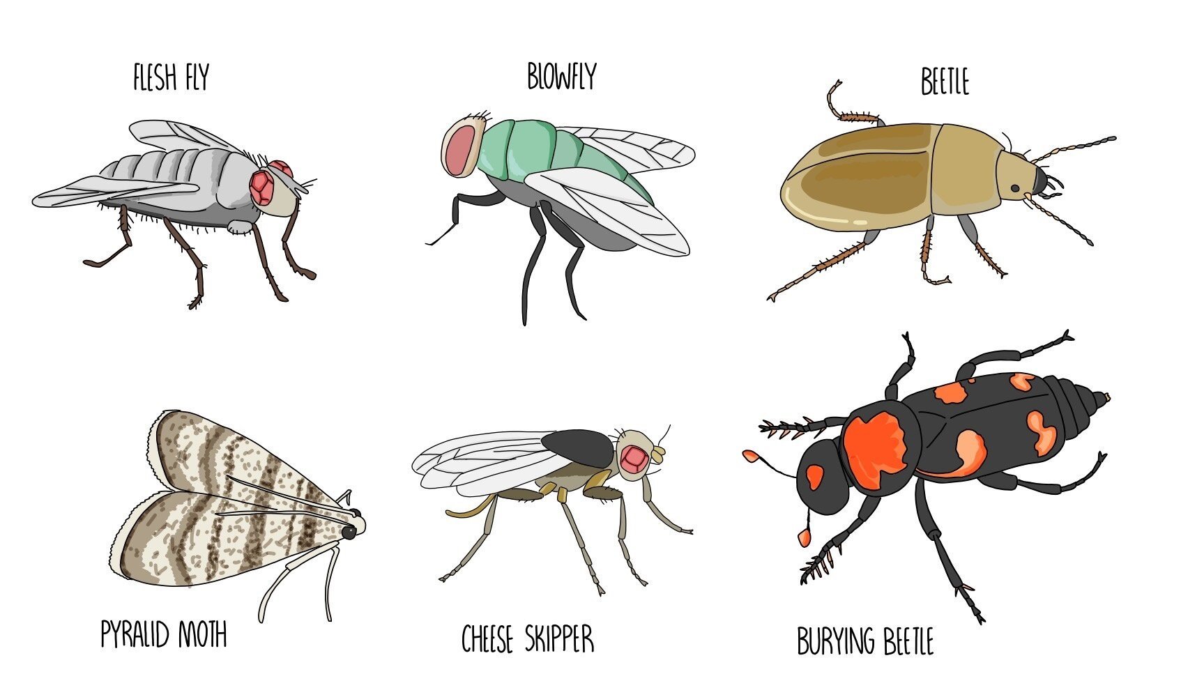
Forensic entomology is the study of the insects feeding and living off a corpse. Depending on the species that is present (and the stage of the life cycle it is in), a time of death can be estimated.
From a few hours - flies are the first insects to appear on the body (blowflies then flesh flies). They will start to lay eggs which take about 24 hours to hatch. This means that if larvae are found on the body, we know that the TOD was less than 24 hours ago.
The next insect to colonise is beetles, which eat decomposed body fat.
A type of moth known as the pyralid moth will appear at a similar time to beetles. They will feed on liquefied parts of the corpse as well as any clothes the body may be wearing.
The next insect to appear is one called the cheese skipper which appears once the body’s proteins have been digested. They feed off digested food molecules and can also feed on any food present in the digestive system which has not been fully digested.
One of the last insects to appear is the burying beetle, which mostly eats dead flesh.
Stage of succession
The type of organism which can be found on a body change over time - this is known as succession. It is similar to plant succession except that most of the early insects, such as flies and beetles, will remain on the body even as new insect species arrive. The stage of succession will be affected by things such as the location of a body - different stages of succession will be seen depending on whether the body is in open air or submerged in water. Succession of a body happens in the following stages:
Bacteria are the first type of organism present on the human body - immediately after death the conditions in a dead body are most favourable to bacteria
As bacteria decompose and digest body tissues, the conditions become more favourable for flies. The flies will lay eggs in the body which will hatch into larvae.
The fly larvae break down tissues of the corpse further, resulting in conditions which are suitable for beetles.
The body begins to dry out, which results in flies leaving the body as they prefer more moist environments. Beetles remain on the body since they can digest dry tissue.
When the body has been fully decomposed, there is no material for the insects to feed on so any remaining insects will leave.
Body temperature
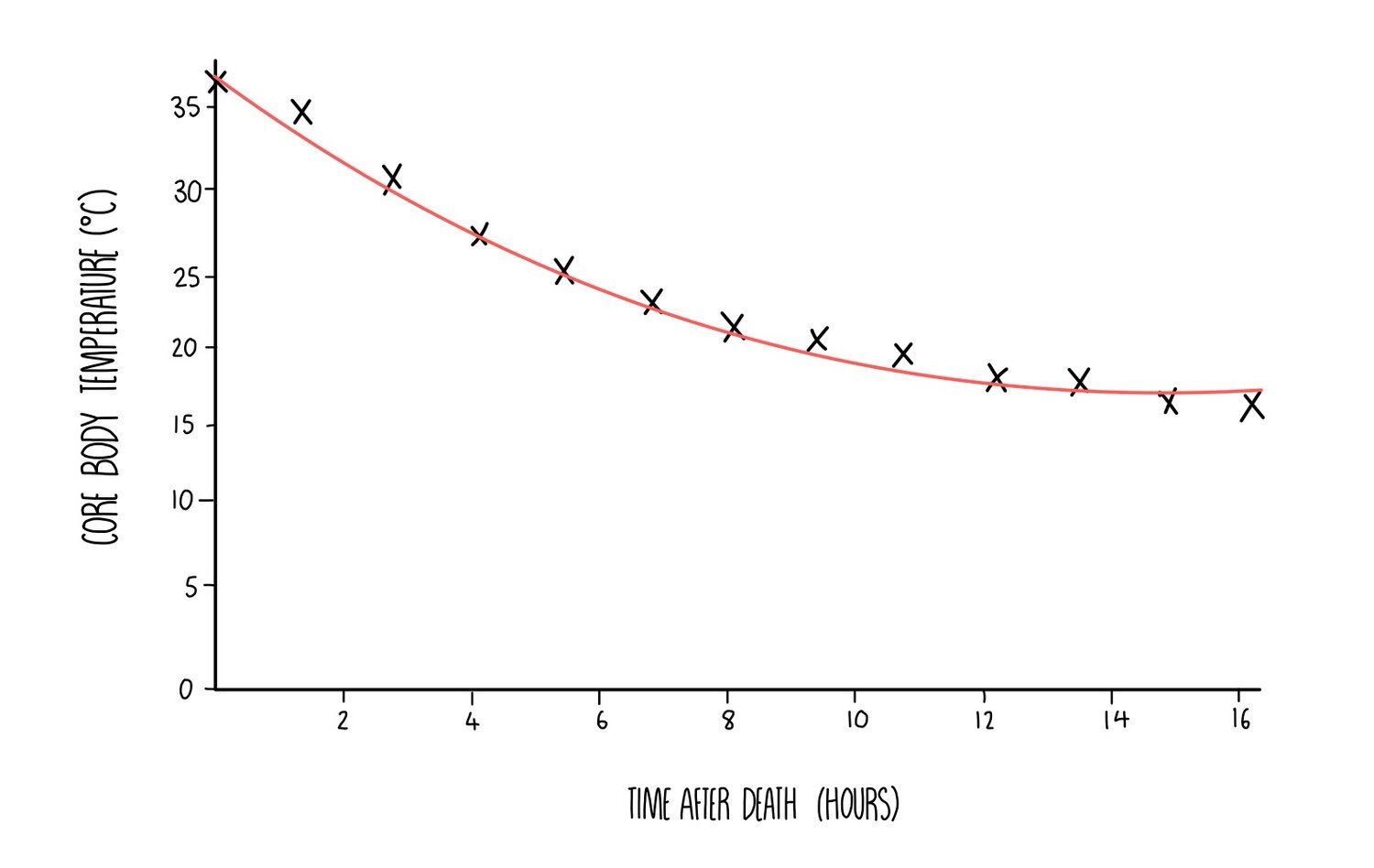
Respiration inside of our cells generates heat as a by-product, maintaining our body temperature at 37oC. When somebody dies, chemical reactions such as respiration stop and their body temperature goes down. Body temperature decreases until there is no difference between the temperature of the body and its surroundings. This reduction in body temperature is known as algor mortis.
Since body temperature decreases at a rate of 1.5oC to 2.0oC each hour after death, the TOD can be calculated by recording the temperature of the corpse. For example, if the temperature of the body is 30oC, we know that it must have been dead for around 3.5 - 4.5 hours. Factors such as the weather, the amount of clothing and the amount of body fat on a person can influence the rate of cooling, so this needs to be taken into account when estimating TOD.
Degree of muscle contraction
Approximately 4-6 hours after death, the body will begin to stiffen in a process called rigor mortis. This happens because oxygen is no longer available since the person has stopped breathing. This means that the muscle cells can no longer respire aerobically so they carry out anaerobic respiration instead, resulting in an accumulation of lactic acid. Lactic acid causes a drop in pH which causes enzymes (such as ATP synthase) to denature. When ATP synthase loses the shape of its active site, it can no longer produce ATP for muscle contraction, resulting in muscle fibres becoming fixed in position and the body stiffens.
Rigor mortis begins with the smaller muscles in the head and ends with the largest muscles in the lower part of the body. By 12-18 hours after death, every muscle in the body will have stiffened. By determining which muscles have undergone rigor mortis, the forensic scientist can calculate an approximate TOD. Factors such as the degree of muscle development and temperature will affect the rate of rigor mortis.
DNA profiling
DNA profiling is a technique which can be used to analyse a sample of DNA (e.g. one found at a crime scene) and compare it to DNA samples taken from the suspects.
The DNA sample will be collected (this is usually blood, saliva or semen) and amplified using PCR.
The PCR products are separated using gel electrophoresis, which separates the DNA fragments according to length.
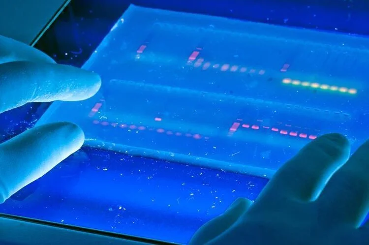
The gel is visualised using UV light and the banding patterns from the suspect’s DNA can be compared with that found at the crime scene.
The same technique can also be used to identify genetic relationships between people (as in paternity testing) or to determine evolutionary relationships between organisms.
Polymerase chain reaction (PCR)
PCR is a technique used to amplify fragments of DNA. This is important because the DNA sample collected at a crime scene doesn’t contain enough material for accurate analysis. Before PCR is carried out, you need to prepare a mixture containing the following:
DNA sample
Free DNA nucleotides
Primers (these are short pieces of DNA which bind to the beginning of the DNA fragment and initiate replication)
The enzyme DNA polymerase which has been extracted from thermophilic (heat-loving) bacteria.
PCR is then carried out in the following stages:
- Separation of the DNA strands - the mixture is heated to 95oC which causes hydrogen bonds between the DNA strands to break.
- Annealing of the primer - the mixture is then cooled to around 60oC which allows the primer to anneal to the DNA.
- DNA synthesis - The temperature is increased to 72oC which is the optimum temperature for DNA polymerase. DNA polymerase forms a new DNA strand from catalysing the formation of phosphodiester bonds between the free DNA nucleotides which align along the DNA template strand by complementary base pairing rules.
Around 30-40 cycles of PCR are carried out, which generates millions of DNA fragments. Each PCR cycle doubles the amount of DNA, so huge numbers of DNA fragments can be quickly generated.
DNA stained with ethidium bromide is separated on by gel electrophoresis. Image: School of Natural Resources from Ann Arbor – CC-BY 2.0
Fluorescent tags
So that the DNA can be visualised, we add a fluorescent molecule which binds to the DNA and makes it visible when exposed to UV light. A common fluorescent tag is ethidium bromide, which inserts itself between the DNA bases and gives off fluorescence under UV light.
Electrophoresis
Once the DNA fragments have been amplified and stained with a fluorescent dye, they need to be separated. This is done using gel electrophoresis, which separates the DNA strands according to length. It works because DNA is negatively charged, which means it will move towards a positive charge when placed in an electric field. Shorter DNA fragments travel through the gel more quickly, which means they will travel a longer distance than the larger DNA fragments.
Gel electrophoresis is carried out in the following steps:
An agarose gel is prepared which contains a row of wells at the top of the gel. The gel is placed into a tank containing buffer solution which is able to conduct electricity.
The DNA sample is mixed with a loading dye - this turns the DNA mixture a dark colour and helps you see what you’re doing. A fixed volume of the DNA samples are pipetted into the wells.
An electrical current is passed through the gel and the DNA will begin to move towards the bottom of the gel (towards the anode).
Once the dye has reached the bottom, the electricity is turned off and the banding pattern is visualised under UV light.


