Gas Exchange
Surface area to volume ratio
For aerobic respiration, organisms need to get oxygen and glucose to their cells and get rid of waste products (carbon dioxide and water). To maintain body temperature, organisms also need to transport heat.
Single-celled organisms like bacteria have a large surface area; a thin surface and a short diffusion pathway so do not need transport systems. They absorb gases by diffusion.
For large, multicellular organisms, their surface area: volume ratio is too small to get these substances by diffusion as there is less space for diffusion to take place.
Due to their size, it would take also too long for the substances to reach cells in the body’s centre (large diffusion pathway).
Therefore, multicellular organisms have mass transport systems (e.g. the circulatory system) and exchange organs (the lungs/intestine).
Adaptations to control body temperature
Animals with smaller surface area to volume ratios lose less heat than ones with more surface area relative to their volume. This explains why smaller organisms have higher metabolic rates – they need to generate more heat energy to replace the heat lost.
Animals adapted to live in hotter climates will have features which increase their surface area. E.g. the desert fox (Fennec fox) has huge ears which stick out to maximise heat loss.
Organisms with a high surface area to volume ratio tend to lose more water by evaporation from their body’s surface. Their kidney is adapted to conserve water e.g. a longer loop of Henle.
Animals adapted to live in cold climates often have thick layers of insulating fat, fur and may hibernate during the coldest months.
Adaptations of exchange surfaces
Most gas exchange surfaces are extremely thin (sometimes just one cell thick), ensuring a short diffusion pathway across the exchange surface. They will also have a large surface area to volume ratio which provides more space for the diffusion of gases. The organism itself will also have features which maximise the concentration gradient of gases across the exchange surface.
Gas exchange in fish

Water (containing oxygen) enters fish through the mouth and pass over the gills. Each gill is made up of several gill filaments stacked up on top of each other. The gill filaments are covered in lamellae to further increase their surface area. The lamellae have very thin walls to reduce the diffusion distance and are filled with capillaries.
Blood in the capillaries flows in the opposite direction to the water to maintain the concentration gradient of oxygen in the blood – this is known as the counter-current system.

Gas exchange in insects
Insects have a tracheal system for gas exchange. Air enters the insect through pores in their outer surface called spiracles. Air then moves down the trachea, which branches off into a large number of tracheoles. The walls of the tracheoles are thin and porous, speeding up diffusion of gases to cells. Rhythmic abdominal movements push air into and out of the spiracles and maintain a steep concentration gradient.
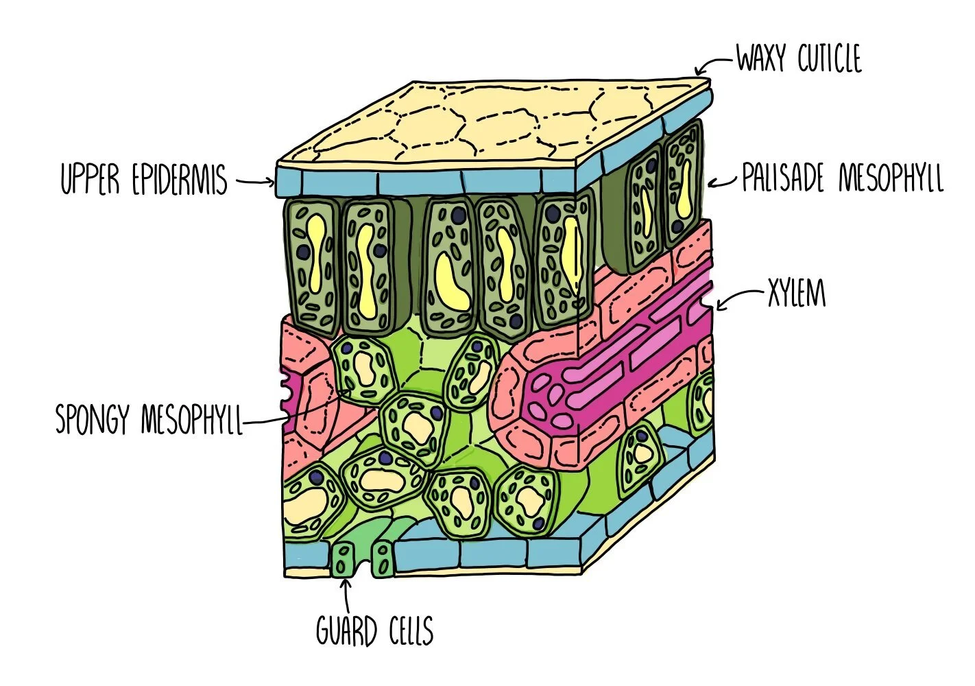
Gas exchange in plants
Plants need to absorb both oxygen (for respiration) and carbon dioxide (for photosynthesis). They also need to get rid of any excess waste gases. Gas exchange in plants happens through the stomata, the holes found in the lower and upper epidermis. Guard cells control the opening and closing of the stomata to prevent excess transpiration. Air spaces in the spongy mesophyll layer allow gases to circulate. Oxygen and carbon dioxide diffuse from these air spaces into the plant cells.
Adaptations to prevent water loss
Insects minimise water loss by:
Closing their spiracles if they become dehydrated
Spiracles are surrounded by small hairs to trap water vapour and reduce the water potential gradient
Covered by a waterproof, waxy cuticle
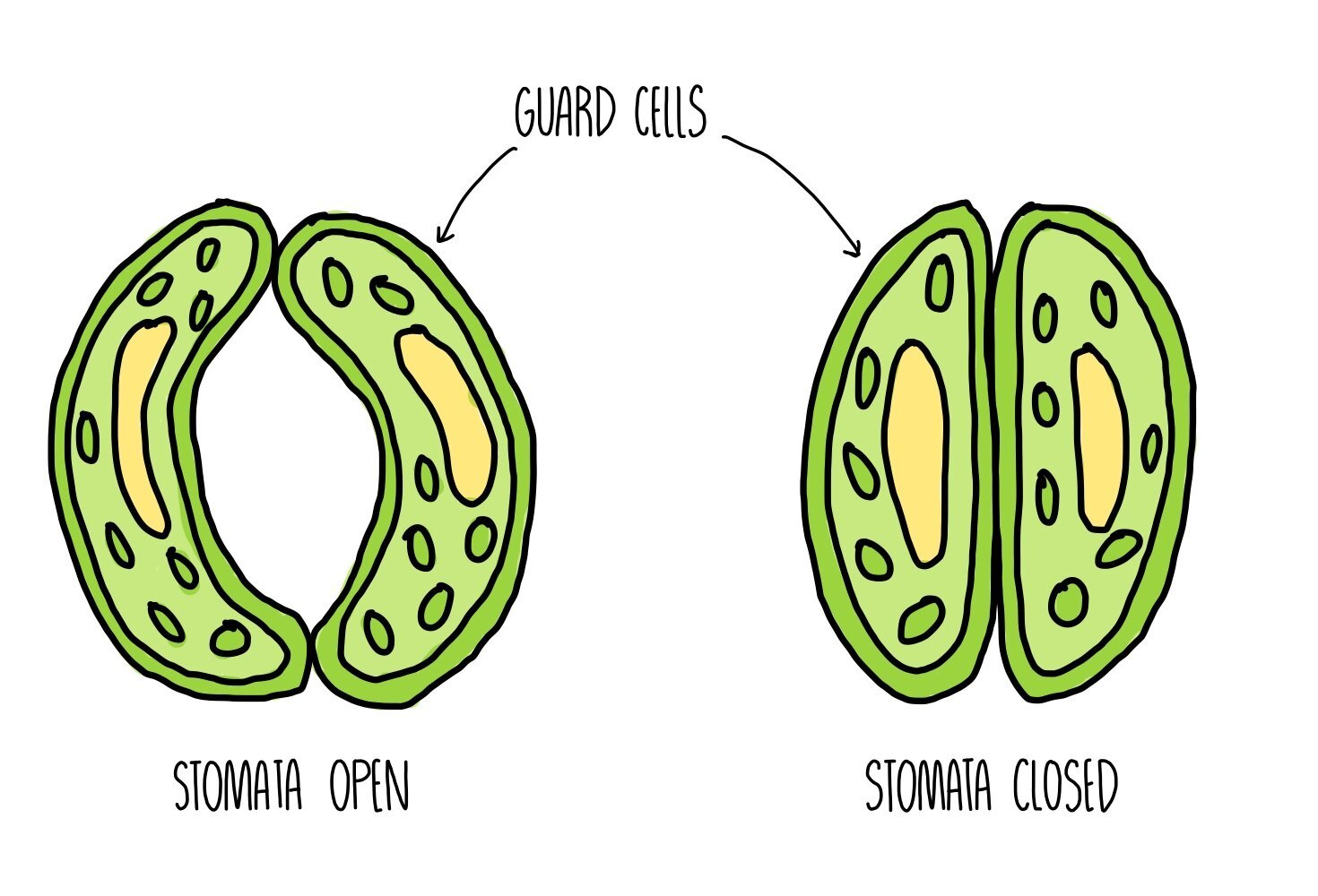
Plants minimise water loss by closing their stomata at night. Water moves out of the guard cells by osmosis, making the guard cells flaccid and closing the stomatal pore.
Plants that are adapted to living in hot, dry conditions are called xerophytes. They have several xerophytic adaptations to prevent excess water loss:
Sunken stomata – stomata are sunken in pits which trap water vapour and reduce the water potential gradient between the inside and outside of the leaf
Hairs around stomata – hairs trap water vapour and reduce the water potential gradient
Curled leaves – also traps water vapour to reduce the gradient
Fewer stomata – so less sites for loss of water
Thicker cuticle – acts as a barrier to evaporation
Respiratory system
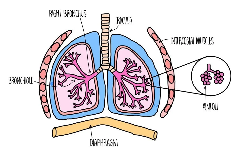
When we breathe, air enters our bodies through our nose and mouth and makes its way down the trachea. The trachea branches into two smaller tubes, called bronchi, which send air to each lung. The bronchi divide into even smaller tubes called bronchioles which finally send the air into air-sacs called alveoli. The alveoli look like a bunch of grapes and it’s this structure where gas exchange takes place. Oxygen diffuses from a region of high concentration in the alveoli to a region of low concentration in the bloodstream, where it travels to different tissues of the body and is used for respiration. Carbon dioxide travels in the other direction, from a region of high concentration in the bloodstream to a region of low concentration in the alveoli, where it travels up the trachea and is breathed out.
The lungs themselves are surrounded by the pleural membrane, a moist membrane which forms an airtight seal around the lungs. The ribcage protects the organs of the respiratory system and are surrounded by intercostal muscles and diaphragm, which move the rib cage during breathing to help bring air into or out of the lungs.
Inhalation and exhalation
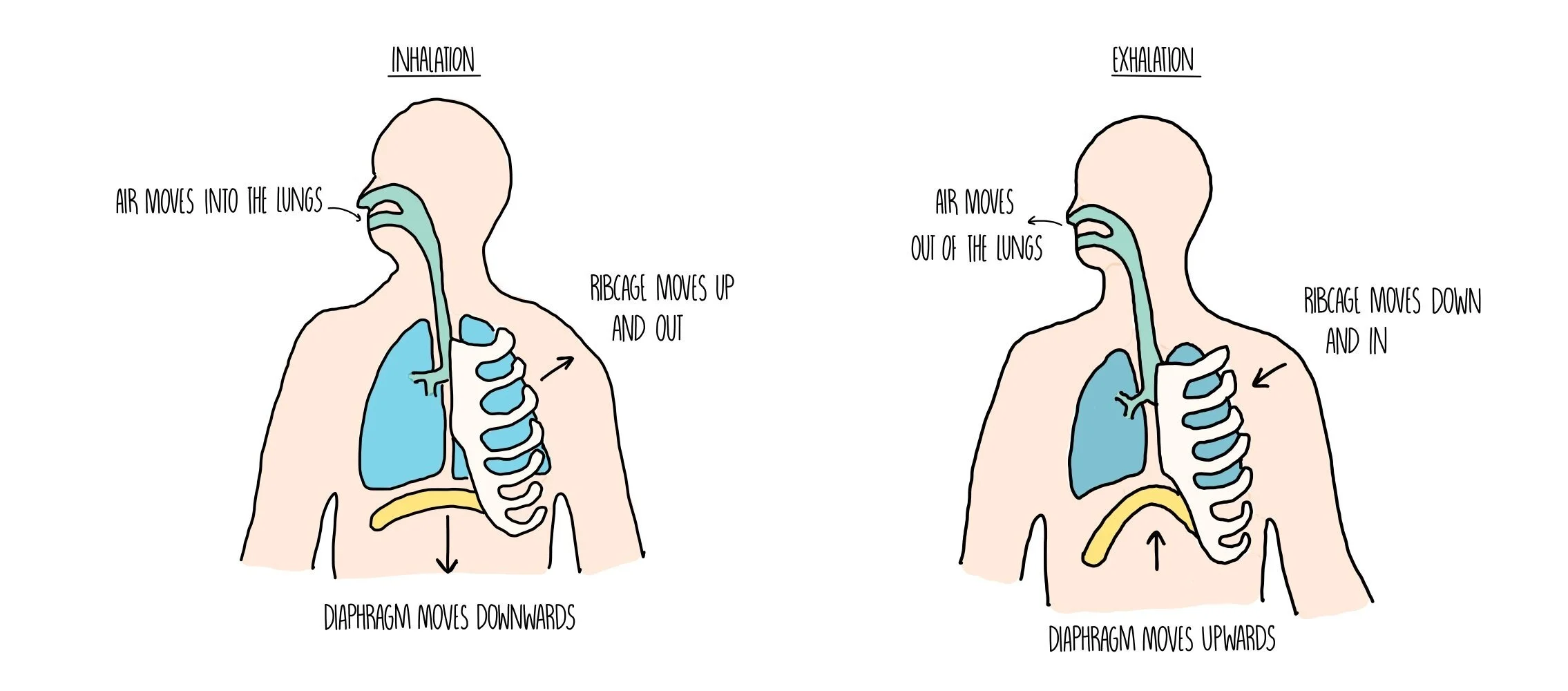
During inhalation, the diaphragm contracts and flattens, while the rib cage moves up and out. This increases the volume of the thorax, resulting in a drop in pressure. The reduced pressure causes air to be sucked into our lungs.
During exhalation, the diaphragm relaxes and forms a dome shape, while the rib cage moves down and in. This reduces the volume of the thorax and increases the pressure, pushing air out of our lungs.
Adaptations of the alveoli
Each alveolus is adapted to make gas exchange as efficient as possible:
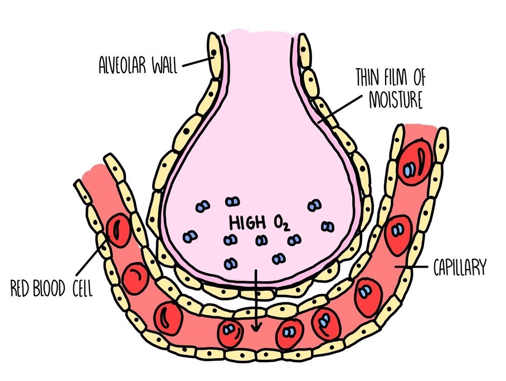
Large surface area: there are approximately 700 million alveoli in our lungs with a combined surface area of 70 square meters.
Good blood supply: lots of capillaries surround each alveolus
Short diffusion distance: the walls of both the alveoli and capillaries are just one cell thick
Moist surfaces: the liquid on the surface of alveoli dissolves gases and facilitates diffusion
Steep concentration gradient: there is always a high concentration of oxygen and carbon dioxide between the alveoli and capillaries. This is maintained by blood flow and ventilation.
Pathway of an oxygen molecule
To get from the inside of an alveolus into the bloodstream, an oxygen molecule has to pass through three cells:
Alveolar epithelium – the single layer of cells that form the alveolar walls
Capillary endothelium – the single layer of cells that form the capillary walls
Red blood cell – once inside the red blood cell, the oxygen molecule binds to a haem group on haemoglobin
Investigating lung function
You can investigate the volume of air that an individual is capable of breathing in and out using a piece of equipment called a spirometer. A spirometer consists of a chamber filled with oxygen and has a lid which moves up and down as a person breathes in and out. The spirometer is attached to a pen, which moves whenever the lid moves to draw a spirometer trace. A spirometer trace can be used to find the following information:
Tidal volume - this is the volume of air in each breath
Forced vital capacity (FVC) - the maximum volume of air that a person can expel from the lungs after a maximum inhalation
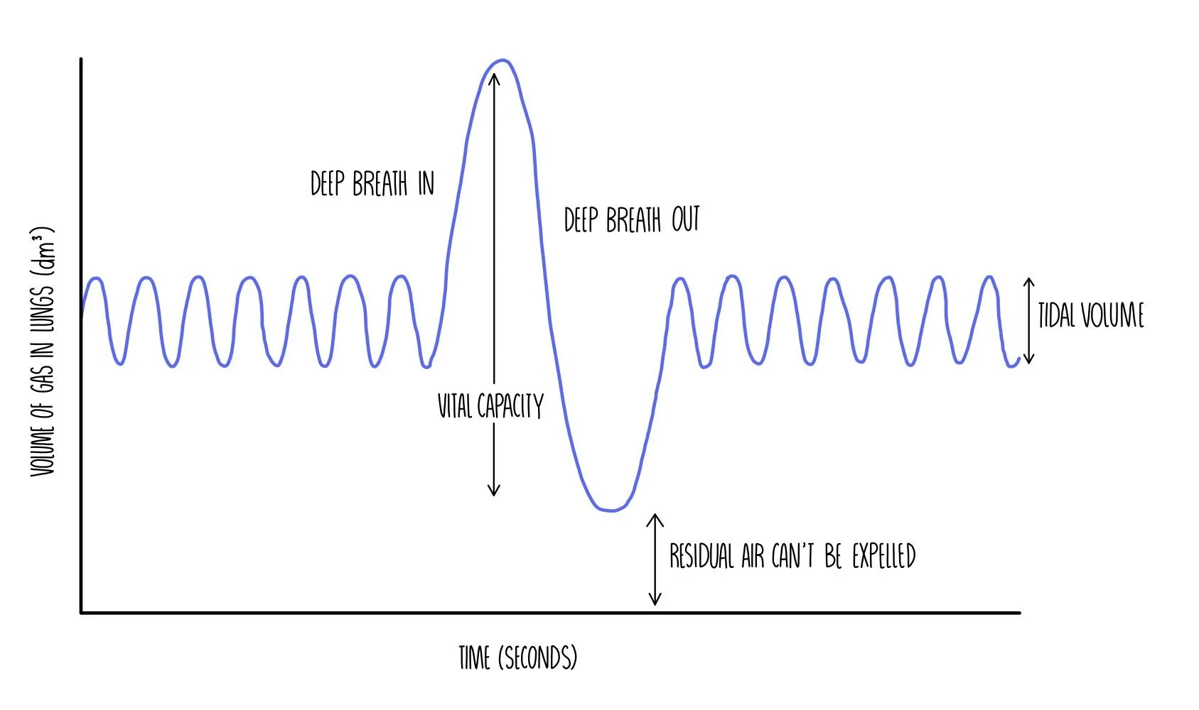
It can also be used to calculate the following:
Breathing rate (aka ventilation rate) - the number of breaths taken per minute
Oxygen consumption - the volume of oxygen used by the body
Forced expiratory volume (FEV1) – the maximum volume of air breathed out in one second
Lung diseases
TB, fibrosis, emphysema and asthma are examples of lung diseases. They all involve impaired gas exchange, so less oxygen moves from the lungs into the bloodstream. Since less oxygen is delivered to cells, less energy is released by aerobic respiration and the patient experiences fatigue.
Tuberculosis
Caused by infection with Mycobacterium tuberculosis bacteria.
Immune cells build a wall around the bacteria, forming hard lumps called tubercles.
Death of tissue in the tubercles damages the alveoli and reduces tidal volume.
Formation of scar tissue (fibrosis) also reduces tidal volume.
Ventilation rate increases to compensate for reduced tidal volume.
Symptoms: cough, chest pain, tiredness, shortness of breath.
Fibrosis
Formation of scar tissue due to infection or exposure to irritants.
Compared to normal tissue, scar tissue is thicker and less elastic.
Less elasticity reduces tidal volume and forced vital capacity (FVC).
Thicker scar tissue increases the diffusion distance, so gas exchange is less efficient.
Ventilation rate increases to compensate for reduced tidal volume.
Symptoms: cough, chest pair, tiredness, and shortness of breath.
Emphysema
Smoking or exposure to pollution causes inflammation of alveoli.
Phagocytes arrive at site of inflammation and produce the enzyme elastase.
Elastase digests elastin within the alveolar walls.
Alveoli lose their elasticity and cannot recoil properly to push air out.
Degradation of alveoli walls results in less efficient gas exchange.
Ventilation rate increases to compensate for reduced tidal volume.
Symptoms: shortness of breath, wheezing.
Asthma
Inflammation of the airways due to an allergic reaction to pollen or dust.
Contraction of smooth muscle in the bronchioles constricts airways and reduces the amount of air breathed out in one second (reduced FEV1).
Symptoms: shortness of breath, wheezing, tight chest.
Drugs delivered by inhalers can relieve symptoms by stimulating the smooth muscle in the bronchioles to relax.