Meiosis and Genetic Variation
Gametes
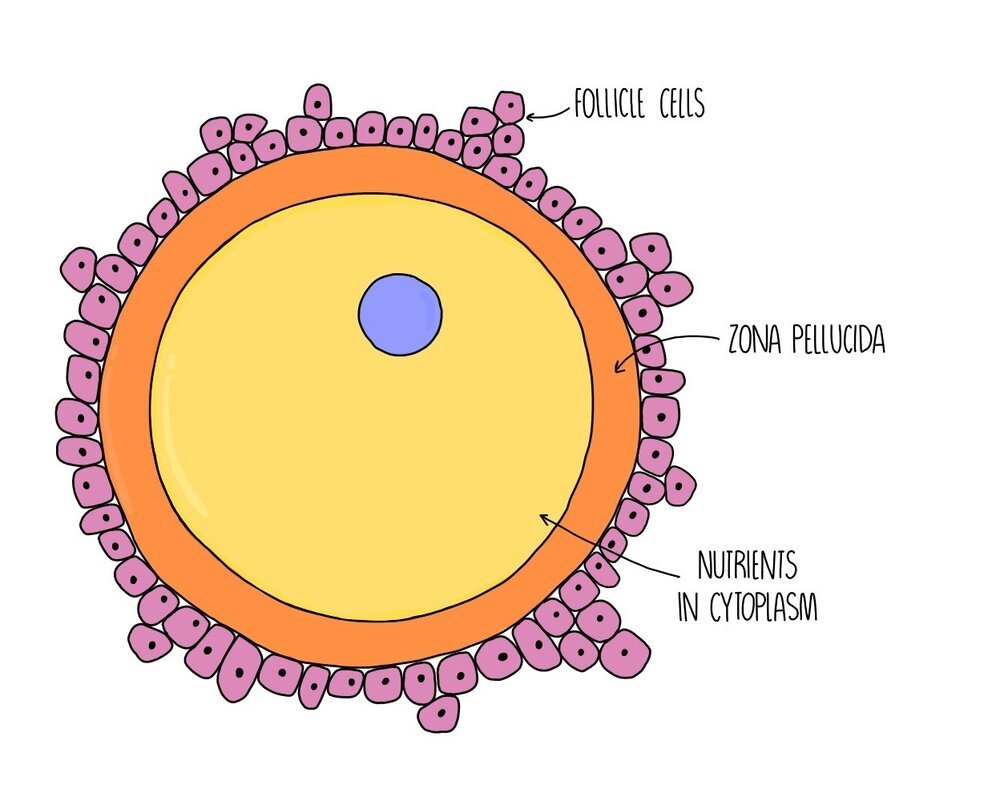
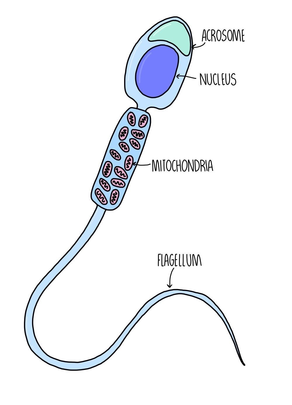
Gametes are sex cells (the sperm and egg in humans). Gametes are haploid which means they contain half the number of chromosomes as the rest of the cells which make up our body. This means that when two gametes fuse during sexual reproduction, the fertilised egg (called a zygote) contains the full number of chromosomes i.e. it is diploid. In humans, the diploid number of chromosomes is 46 (23 pairs), which means that gametes contain just 23 chromosomes.
During sexual reproduction, the nucleus of the sperm cell fuses with the nucleus of the egg cell - this fusion of nuclei is called fertilisation.
Meiosis
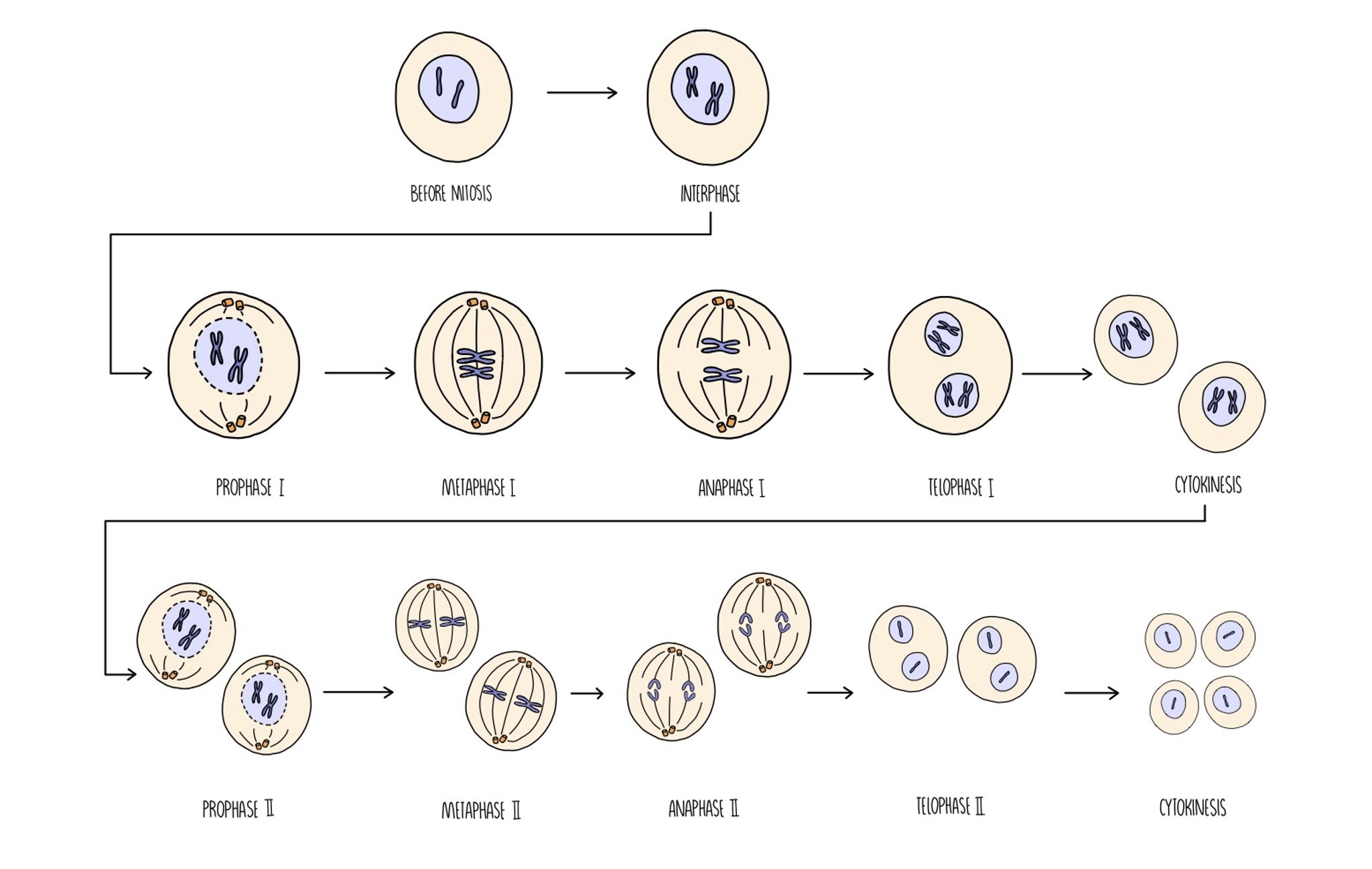
Meiosis is the type of cell division which produces gametes for sexual reproduction. Unlike mitosis, the daughter cells are genetically different from the parent cell and contain just half the number of chromosomes (i.e. they are haploid). When two haploid gametes join during fertilisation, a diploid cell called a zygote is formed. Meiosis involves two rounds of cell division which are referred to as meiosis I and meiosis II.
It takes place in the following stages:
Meiosis I
Interphase: the DNA replicates so there are now two identical copies of each chromosome (referred to as chromatids).
Prophase I: chromatids condense and arrange themselves into homologous pairs (called bivalents). Crossing over occurs (see below). The nuclear envelope disintegrates and spindle fibres form.
Metaphase I: homologous chromosomes line up along the equator and attach to the spindle fibre by their centromeres.
Anaphase I: homologous chromosomes are separated
Telophase I: chromosomes reach opposite poles of the cell. Nuclear envelope reforms around the chromosomes. Cytokinesis results in the formation of two daughter cells.
Meiosis II
Prophase II: chromosomes condense, nuclear envelope disintegrates and spindle fibres form.
Metaphase II: chromosomes attach to the spindle fibre by their centromeres.
Anaphase II: sister chromatids are separated.
Telophase II: chromatids reach opposite poles of the cell. Nuclear envelope reforms and cytokinesis takes places. Four genetically unique daughter cells are produced.
Meiosis increases genetic variation
From an evolutionary point of view, it is important that organisms produce offspring that show as much genetic variation as possible. Imagine if a mother duck gave birth to a group of ducklings that were all had very similar genes - these ducklings will all be equally vulnerable to the same diseases and other threats to their survival. Meiosis increases genetic variation in two ways - crossing over and independent assortment.
Crossing Over
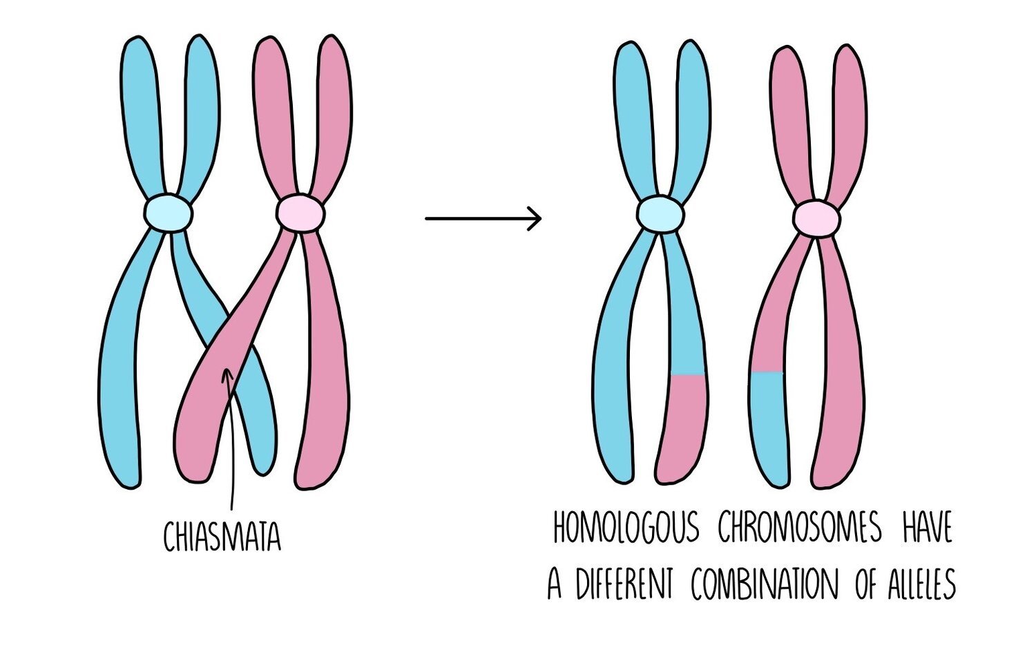
During prophase I of meiosis, a process called crossing over occurs. This is when the homologous chromosomes move towards each other and exchange genetic material. A chromatid from the maternal chromosome becomes twisted around the paternal chromosome and they connect through a structure called the chiasmata. Pieces of chromosomes are exchanged and the chromatids separate, forming chromosomes with different combinations of alleles.
Independent assortment
Depending on the order in which chromosomes line up along the equator of the cell during metaphase, different combinations of chromosomes will end up in each gamete. The way in which the chromosomes align themselves on the spindle fibre is completely random, resulting in a huge number of possibilities of chromosomal combinations in the gametes.
Chromosome mutations
Errors in meiosis can sometimes result in gametes ending up with the wrong number of chromosomes e.g. a gamete may have a chromosome missing, gain an extra chromosome or extra bits of chromosome. These chromosomal mutations are inherited by the next generation because the errors are present in the gametes.
Down’s syndrome is an example of a disorder caused by a chromosomal mutation. People with Down’s syndrome have an extra copy of chromosome 21 because the homologous chromosomes failed to separate in anaphase I. Failure of homologous chromosomes to separate is known as non-disjunction.
Nucleotide mutations
A mutation is a change to the base sequence of DNA occurring when the cell makes errors during DNA replication. Since the order of bases within a gene determine the amino acid sequence (or primary structure) of a protein, a mutation can result in an altered primary structure. Primary structure then determines later stages of folding, so a mutation can result in a change to the overall 3D structure (tertiary structure) of a protein. This could result in the protein losing its function.
Types of mutation:
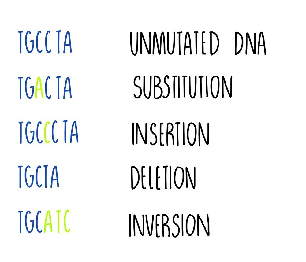
Substitution - one base is replaced for another e.g. TGCCTA becomes TGACTA. It results either in a change to a single amino acid, or the amino acid might stay the same because more than one codon can code for the same amino acid (the genetic code is degenerate).
Insertion - one or more bases is added e.g. TGCCTA becomes TGCCCTA. This type of mutation will change the codon (and perhaps the amino acid) and all following codons (this is called a frameshift).
Deletion - one or more bases is removed e.g. TGCCTA becomes TGCTA. Deletion mutations will change the codon at the point of mutation and all following codons, resulting in a frameshift.
Inversion - a sequence of bases is reversed e.g. TGCCTA becomes TGCATC. It will result in a change to a single amino acid.
Some mutations can have a neutral effect on a protein’s function. This could be because:
The mutation changes a base in a triplet but the amino acid that the triplet codes for doesn’t change. This happens because some amino acids are coded for by more than one triplet (the genetic code is degenerate).
The mutation produces a triplet that codes for a different amino acid but the amino acid is chemically similar to the original so it functions like the original amino acid.
The mutated triplet codes for an amino acid not involved with the protein’s function e.g. one that is located far away from an enzyme’s active site, so the protein works as it normally does.
However, some mutations will have an effect and it could be beneficial or harmful to the organism. An example of a beneficial mutation is antibiotic resistance in bacteria (from the bacteria’s perspective). The mutation would allow it to survive in the presence of the antibiotic whereas it would have previously been killed. Other mutations will have harmful effects, such as the mutations which cause cystic fibrosis or cancer.
Mutations are random events but the rate of mutation can increase if cells are exposed to mutagenic agents – things like UV radiation, the chemicals in cigarette smoke and certain viruses.
