Mutations and Gene Expression
Types of mutation
Any change to the base sequence of DNA is called a mutation. There are various types:
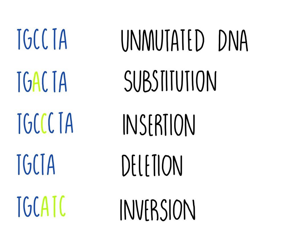
Substitution - one base is replaced for another e.g. TGCCTA becomes TGACTA. It results either in a change to a single amino acid, or the amino acid might stay the same because more than one codon can code for the same amino acid (the genetic code is degenerate).
Insertion - one or more bases is added e.g. TGCCTA becomes TGCCCTA. This type of mutation will change the codon (and perhaps the amino acid) and all following codons (this is called a frameshift).
Deletion - one or more bases is removed e.g. TGCCTA becomes TGCTA. Deletion mutations will change the codon at the point of mutation and all following codons, resulting in a frameshift.
Inversion - a sequence of bases is reversed e.g. TGCCTA becomes TGCATC. It will result in a change to a single amino acid.
Some mutations can have a neutral effect on a protein’s function. This could be because:
The mutation changes a base in a triplet but the amino acid that the triplet codes for doesn’t change. This happens because some amino acids are coded for by more than one triplet (the genetic code is degenerate).
The mutation produces a triplet that codes for a different amino acid but the amino acid is chemically similar to the original so it functions like the original amino acid.
The mutated triplet codes for an amino acid not involved with the protein’s function e.g. one that is located far away from an enzyme’s active site, so the protein works as it normally does.
However, some mutations will have an effect and it could be beneficial or harmful to the organism. In these cases, the mutated allele produces a protein with an altered tertiary structure, due to changes in protein’s primary structure (i.e. a different order of amino acids).
An example of a beneficial mutation is antibiotic resistance in bacteria (from the bacteria’s perspective). The mutation would allow it to survive in the presence of the antibiotic whereas it would have previously been killed. Other mutations will have harmful effects, such as the mutations which cause cystic fibrosis or cancer.
Causes of mutation
Mutations happen randomly due to errors during DNA replication. But harmful things in the environment (mutagenic agents) can make mutations more likely. Examples of mutagens include ionising radiation, UV radiations and certain chemicals, such as the chemicals found in pesticides. They work in different ways:
Some chemicals delete or alter bases. For example, alkylating agents changes the structure of guanine so that it pairs with thymine rather than cytosine.
Some types of radiation change DNA structure. For example, UV radiation causes neighbouring thymines to join together
Some chemicals are a similar shape to bases and can become incorporated into the DNA strand during replication. For example, 5-bromouracil can substitute for thymine, pairing with guanine instead of adenine and triggering a substitution mutation in the DNA.
Cancer
Individuals acquire mutations throughout their life. If a mutation occurs in a gene that controls the rate of cell division, it can lead to the formation of a tumour. A tumour is an abnormal mass of cells which develops into a cancer if they begin to invade and destroy surrounding tissue.
Mutations in two types of genes can lead to cancer:
Mutations which inactivate tumour-suppressor genes – these genes stop uncontrolled cell division by coding for proteins which inhibit mitosis or trigger apoptosis. If the tumour suppressor gene is inactivated, the protein isn’t produced, resulting in uncontrolled cell division.
Mutations which activate proto-oncogenes – these genes stimulate cell divisions by coding for proteins which trigger mitosis. If the proto-oncogene is overactive, cells divide uncontrollable, leading to the formation of a tumour.
Tumours can either be benign or malignant:
Benign tumours are non-cancerous and grow slowly. They’re covered in fibrous tissue that stops them from invading other parts of the body. They are not usually harmful, although they can cause blockages and put pressure on organs. Some benign tumours become malignant.
Malignant tumours are cancerous. They grow quickly and spread around the body, invading multiple tissues.
Compared to normal cells, cancer cells have some distinct features:
Express different antigens on their cell membrane
Larger, darker nuclei
Divide by mitosis more frequently
Irregular shape
Don’t produce all the proteins that a cell needs to function properly
Don’t respond to growth-regulating processes
Epigenetics
Epigenetic modification involves the addition or removal of chemical ‘tags’ onto DNA or histone proteins. These modifications are caused by environmental factors (such as diet, stress and smoking) and can occur from as early as when we are in the womb.
The addition or removal of epigenetic tags changes the structure of the chromosome, making it either more or less accessible to RNA polymerase, transcription factors and other proteins which are involved in transcription. The more open the structure of the chromosome, the more accessible it is to RNA polymerase - the gene is switched on. The less open the structure, the less accessible it is and transcription is reduced - the gene is switched off. The two main types of epigenetic ‘tags’ are methyl groups and acetyl groups.
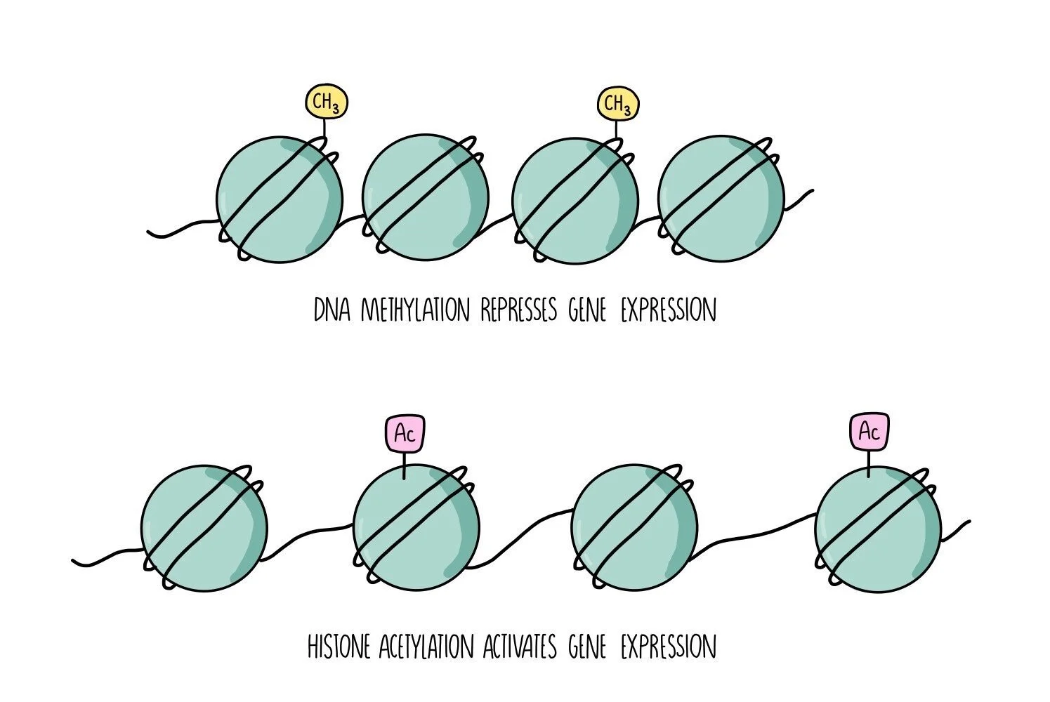
DNA methylation
Methyl groups (-CH3) can be added directly onto the DNA. They attach to a region of DNA called the CpG site - this is where a cytosine is found next to a guanine. The addition of a methyl group makes the DNA less accessible to the proteins involved in transcription. The gene is switched off (inactivated).
Histone acetylation
Chromosomes are made up of a substance called chromatin, which consists of DNA wrapped around histone proteins. Chromatin can either be tightly wound (condensed) or more loosely wound (less condensed). The more condensed the chromatin, the less accessible it is to the proteins involved in transcription. The addition of acetyl groups to histones causes the chromatin to become less condensed, making it more accessible and activating the gene. The removal of acetyl groups converts the chromatin to a more highly condensed form, which is less accessible and represses transcription of the gene. Histone deacetylases (HDAC) enzymes remove acetyl groups from histones and so contribute to reduced gene expression.
Uncontrolled cell division can be caused by increased methylation (hypermethylation) of tumour suppressor genes or reduced methylation (hypomethylation) of proto-oncogenes.
Drugs that reduce DNA methylation are used in chemotherapy for types of cancer that are caused by hypermethylation of tumour suppressor genes. Drugs which inhibit HDACs (the enzymes that remove acetyl groups from histones) are also being used to treat some types of cancer. This prevents deacetylation and allows genes to remain switched on. The problem is that it’s hard to make these drugs specifically target cancer cells while leaving other cells alone.
Role of oestrogen in cancer
Increased exposure to oestrogen (e.g. prolonged treatment with oestrogen-containing contraceptive pills or menopause treatments) is thought to increase the risk of breast cancer. This may be because:
Oestrogen stimulates cell division in breast cells. If cells are dividing more frequently, there is more opportunity for mutation to take place.
If a mutation does happen, oestrogen may help the mutated cells to replicate faster, helping tumours to form quickly.
Oestrogen may even introduce mutations directly into the DNA of breast cells.
Stem Cells and Potency
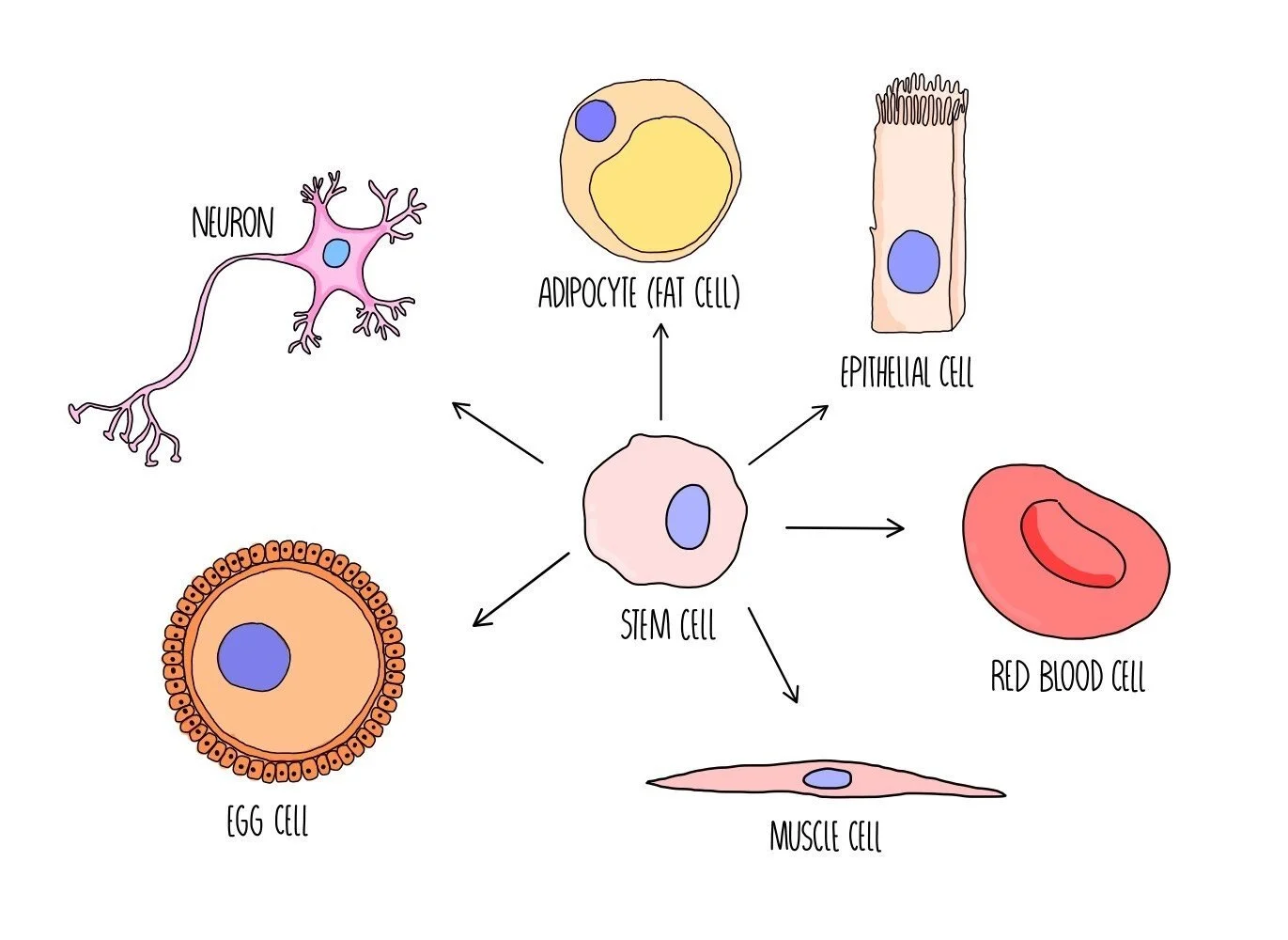
Stem cells are unspecialised cells which have the ability to become specialised cells, such as heart cells or neurons. The process by which a stem cell is converted from an unspecialised cell to a specialised cell is called cell differentiation. Stem cells have an unlimited capacity to divide and can produce lots more stem cells by mitosis.
The ability of stem cells to undergo differentiation is referred to as potency - there are different levels of potency:
Totipotent - totipotent cells have the ability to divide into any type of cell (including the extraembryonic cells which make up the placenta and umbilical cord).
Pluripotent - pluripotent cells can divide into any type of cell except the extraembryonic cells.
Multipotent - these cells can divide into a handful of different cell types
Unipotent - these cells can only divide into one type of cell
Gene Expression
All cells of our body contain exactly the same set of genes but can have very different structures and functions in our body. The reason that a brain cell is so much different from a muscle cell is due to the fact that different proteins are being made - this results from the activation (and deactivation) of different genes in these two cells.
Within each cell, certain genes will be activated and others will be inactivated. Only the activated genes are transcribed into mRNA which is translated into protein. The particular proteins that are formed will modify the cell by changing its structure and controlling cellular processes. These changes cause the cell to become specialised.
For example, during the differentiation of a stem cell into a red blood cell, certain genes are activated. These genes are responsible for the production of haemoglobin and of proteins which will destroy the nucleus (enabling the cell to pack in more haemoglobin). Other genes will be inactivated so that any proteins which are unrelated to the functioning of red blood cells will not be produced. The production of red blood cell-related proteins will modify the cell, resulting in a specialised red blood cell.
Stem cells in medicine
Stem cells are being used as a treatment for certain diseases:
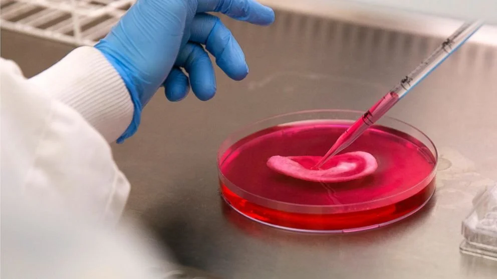
Stem cells are grown on a ear-shaped scaffold. Credit: Matt Dunham (AP Photo)
Stem cell transplants are given to patients with leukaemia - leukaemia is a type of cancer which destroys stem cells so bone marrow transplants are used to replace the lost stem cells.
Research is being carried out to develop ways of growing whole organs from stem cells. The organs can then be transplanted into the patient to replace organs that have been damaged or are diseased e.g. pancreatic transplants can be given to people with diabetes. This approach will help those who currently have to wait years for an organ donation.
Adult stem cells vs embryonic stem cells
There are two sources of stem cells: adult stem cells (ASCs) and embryonic stem cells (ESCs). ASCs are found in the bone marrow of adults and have a more limited potency. They can only develop into a limited number of cell types (i.e they are multipotent). ESCs are found in human embryos and can develop into all types of adult cell (i.e. they are pluripotent). The ability of ESCs to divide into any cell type makes them much more useful for medicine but the use of ESCs comes with ethical implications, since they are obtained from embryos that have developed in the lab from in vitro fertilisation. ESCs are taken from the embryos when they are 4-5 days old and are then discarded. Some people are opposed to this since they believe that a human has a right to life from the moment of conception.
There’s a third type of stem cell – induced pluripotent stem cells (iPS cells). These are made by taking adult stem cells and reprogramming them into a stem cell state using a cocktail of transcription factors. The transcription factors cause the adult cells to express genes associated with pluripotency. The transcription factors are introduced into cells by packaging them inside a virus, which then infects the adult cells.
iPS cells are seen as the most useful type of stem cell in research. They don’t have the ethical baggage that ESCs but can be used to make any cell type (unlike ASCs). They can even be made from a patient’s own cells. This means that the iPS cells are genetically identical to the patient, reducing the risk of rejection.
Transcription Factors
The activation and deactivation of genes is carried out by proteins called transcription factors (TFs). TFs which activate genes are called activators whereas TFs which deactivate genes are referred to as repressors. Activators can work by binding to the beginning of the gene (the promoter region) and helping RNA polymerase to bind and transcribe the gene. Repressors can work by binding to the gene and blocking RNA polymerase from binding.
Oestrogen works by binding to an oestrogen receptor (a transcription factor) to form an oestrogen-oestrogen receptor complex. Oestrogen is a steroid hormone (made of lipid) so it can cross the cell membrane and get inside the nucleus. Once there, it binds to specific sites on DNA, either activating or inhibiting the expression of its target genes.
RNA interference
RNA interference is when small strands of RNA bind to messenger RNA (mRNA) to form a double-stranded structure. This prevents the mRNA from being translated into proteins and prevents the gene from being expressed.
There are two types of interfering RNA – small interfering RNA (siRNA) and microRNA (miRNA). These are just short lengths of RNA that do not code for proteins.
Small interfering RNA:
Double-stranded siRNA binds to proteins in the cytoplasm and unwinds.
A single strand of siRNA binds to the target mRNA – the siRNA will have a complementary sequence to its target so will only bind to specific mRNAs.
The proteins that are bound to the siRNA chop the mRNA into fragments.
The small mRNA fragments are moved into a processing body for degradation.
MicroRNA:
Unlike siRNA, miRNA isn’t completely complementary to its target mRNA so it is less specific.
miRNA binds to proteins in the cytoplasm
The miRNA-protein complex binds to the target mRNA and blocks the ribosome from translating the mRNA.
The mRNA is moved into a processing body and is either stored (and may be translated in the future) or degraded.
