Reproduction
Some organisms can reproduce both sexually and sexually depending on their circumstances. During sexual reproduction, genes from both parents are passed onto offspring. Genes are stretches of DNA which code for proteins, a major component of cells.
Sexual and asexual reproduction
Mitosis and meiosis are both forms of cell division. Mitosis is responsible for producing clones (genetically identical cells) for growth and repair whilst meiosis is responsible for producing genetically unique gametes (sex cells) for sexual reproduction.
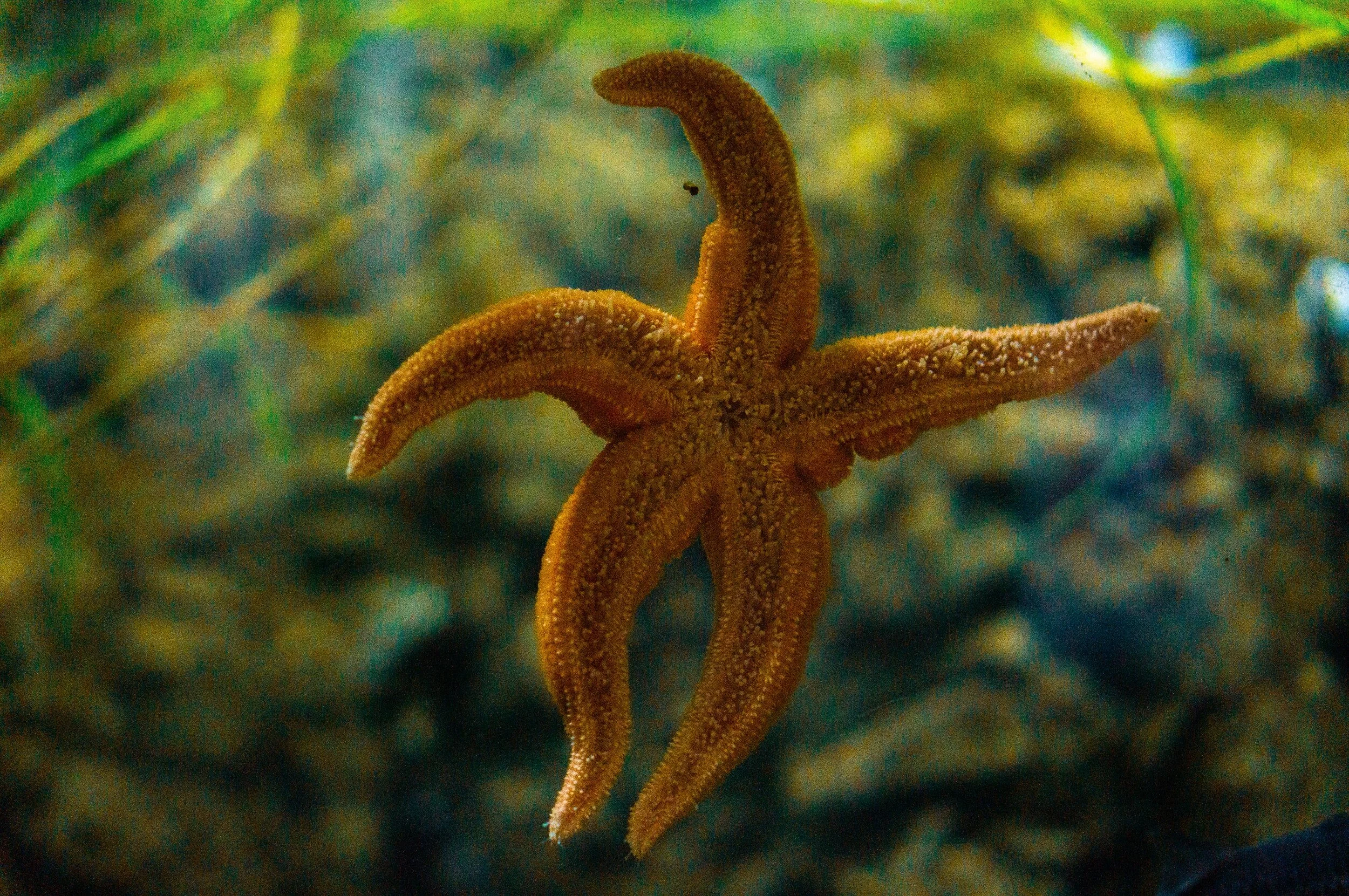
Starfish can reproduce asexually
Sexual reproduction involves the fusion of male and female gametes.
In animals, the male gamete is a sperm cell and the female gamete is an egg cell.
In plants, the male gamete is a pollen grain and the female gamete is an egg cell (ovum).
The two gametes fuse and mix genetic information, leading to variety in the offspring.
Meiosis is the process in which gametes are formed.
In contrast, asexual reproduction:
Does not involve the fusion of gametes — only one parent is involved.
There is no mixing of genetic information so the offspring produced are clones (genetically identical) to the parent.
Organisms which are able to reproduce asexually include starfish, wasps and in rare cases, sharks.
Mitosis is the type of cell division involved in asexual reproduction.
Meiosis
Meiosis is the type of cell division which produces gametes for sexual reproduction. It takes place in the reproductive organs (ovaries and testes). Unlike mitosis, the daughter cells are genetically different from the parent cell and contain just half the number of chromosomes (i.e. they are haploid). Meiosis involves two rounds of cell division which are referred to as meiosis I and meiosis II.
When a cell divides by meiosis:
The DNA replicates
The cell divides twice to form four daughter cells, each with a single set of chromosomes
All gametes are genetically different from each other
When two haploid gametes join during fertilisation, a diploid cell called a zygote is formed. The zygote then divides by mitosis to form a ball of cells which develops into an embryo. The cells in the embryo start to differentiate to become various different body organs, such as the brain and the liver.
Advantages and disadvantages of sexual and asexual reproduction
Sexual and asexual reproduction both have their advantages and disadvantages. Some organisms are able to reproduce both sexually and asexually and will choose one or the other depending on the circumstances they find themselves in.
Advantages of sexual reproduction:
Produces variation in the offspring – this means that all of the offspring aren’t equally vulnerable to things like disease.
Without variation there would be no natural selection. Natural selection is advantageous because it makes organisms more suited (adapted) to their environment.
Selective breeding relies on natural selection to produce organisms with favourable characteristics for food production.
Advantages of asexual reproduction:
Time and energy is not wasted trying to find a mate
Quicker than sexual reproduction
When conditions are favourable, many identical organisms can be produced
Only one parent needed
Gardeners use asexual reproduction to produce lots of plants from a single parent plant with desirable characteristics
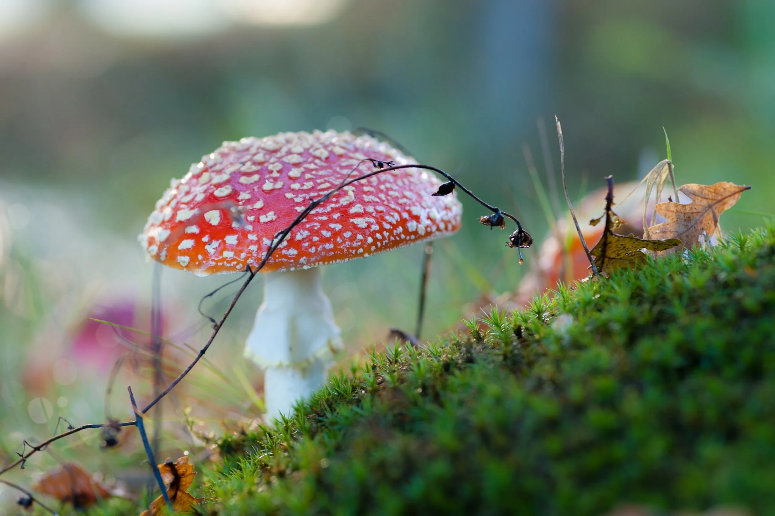
Fungi can reproduce sexually and asexually depending on external conditions.
Some organisms are able to reproduce by both methods, depending on their situation:
The Plasmodium parasite which causes malaria reproduces asexually in humans but reproduces sexually in the mosquito.
Fungi can reproduce both sexually to introduce variation and asexually when they form spores.
Strawberry plants can reproduce sexually but can reproduce asexually by growing runners.
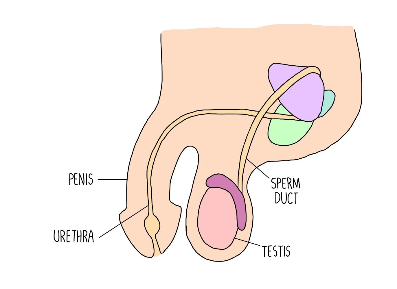
Male reproductive system
Testes (singular: testi) are the site of sperm and testosterone production. Sperm travels along a tube called the sperm duct, where it mixes with seminal fluid which provides the sperm with nutrients. The combination of sperm with the seminal fluid creates semen. During ejaculation, the semen travels along the urethra and out of the body through the penis. The penis is also used to pass urine out of the body, via the urethra.
Female reproductive system
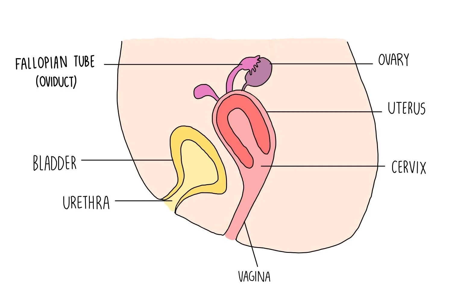
Female gametes are called ova (singular: ovum) which are found in the ovaries. After an egg matures in the ovaries, it is released into the oviduct (fallopian tube) and moves through the tube towards the uterus. The oviduct is lined with ciliated epithelial cells, which have finger-like projections called cilia to move and sweep the ovum along the tube. It is in the oviduct where fertilisation takes place, if the ovum successfully encounters a sperm cell. Once in the uterus, the fertilised egg (zygote) implants itself into the uterine lining where it will develop into an embryo. If no fertilisation occurs, the uterine lining will shed and pass out of the body during menstruation.
A circular ring of muscle called the cervix is found at the end of the uterus and it keeps the baby in position during pregnancy. The vagina leads from the cervix to the outside of the body and it holds the penis during sexual intercourse. The urethra is a tube separate from the vagina, for allowing urine stored in the bladder to pass out of the body.
Pregnancy
If a sperm and egg cell fuses, a fertilised egg (or zygote) is produced. The zygote divides by mitosis into a ball of cells called an embryo which implants itself into the wall of the uterus. The embryo is surrounded by a liquid called amniotic fluid which cushions the baby and acts as a shock-absorber, protecting the embryo. A placenta also forms and is connected to the embryo through the umbilical cord. The placenta anchors the baby in the uterus and provides the developing embryo with substances (such as oxygen and glucose) as well as removing any waste products (like carbon dioxide).
DNA and the genome
A gene is a section of DNA which codes for a protein. Proteins are like molecular ‘machines’ and come in a variety of forms, from antibodies to enzymes. Each protein has its own gene, which provide the cell with instructions on how to make it. Genes are located on chromosomes within the nucleus.
A molecule of DNA is made up of two strands which are twisted around each other to form a double helix.
The entire DNA within an organism is called its genome. The Human Genome Project was set up in 1990 to study the entire human genome. It took scientists 13 years to identify every single base present in human DNA and is a really useful tool in medicine and research. For example:
It has enabled scientists to search for and identify genes which are linked to disease
It has helped scientists understand and treat genetic disorders
It has allowed us to trace human migration patterns in the past
The structure of a nucleotide.
DNA structure
DNA is a polymer made up of long chains of nucleotides bonded together. Each nucleotide consists of a sugar, a phosphate group and a base. The bases present in DNA include adenine (A), thymine (T), guanine (G) and cytosine (C) where A always bonds to T and G always bonds to C. This happens because these pairs fit neatly together and we say their shape is complementary.
DNA and RNA nucleotides form complementary base pairs.
That means that if a DNA molecule consists of 20% guanine then it should also contain 20% cytosine, since there will always be equal amounts of the complementary base. We can then work out that the remaining 60% must be made up of the other two bases, adenine and thymine (30% of each).
For a gene to produce a protein, the DNA within the gene must first be copied into RNA in a process called transcription. This is important because DNA is too big and wrapped snuggly around chromosomes to leave the nucleus. Just like photocopying a single useful page out of a chunky library book, the cell makes an RNA copy of all the important information contained in the gene. RNA is similar to DNA, but has two important differences:
DNA is double-stranded whereas RNA is a single strand
RNA contains uracil in place of thymine
There are lots of different types of RNA which perform different functions. The type we are talking about here, which is used to transfer information from DNA for protein synthesis are called messenger RNA or mRNA. Once the mRNA molecule has been synthesised, it can leave the nucleus and enter the cytoplasm. From there it finds its way to structures called ribosomes, which are basically protein-building machines. The ribosome attaches itself to the RNA and slides along it. The ribosome ‘reads’ the mRNA in a series of three bases (such as AUG, CCA, GCU) called codons. Each codon corresponds to a particular amino acid. As the ribosome reads the codons, a transfer RNA (tRNA) molecule which has a complementary anticodon carries an amino acid to the ribosome. Once the ribosome has read through the length of the mRNA, a series of different amino acids will have been dropped off by several tRNA molecules. Bonds form between the amino acids to form a protein chain. When the protein chain is complete, it folds into a unique shape. This unique shape enables the proteins to do their job as enzymes, hormones or structural components such as collagen.
Because different RNA codons (triplets of bases) code for different amino acids, this means that the sequence of bases in a gene determines the order of amino acids as they are assembled during protein synthesis. If a mutation occurs so that the DNA base changes, this will result in a change in the order of amino acids. This could result in a protein being produced which has a different shape and it may not work as effectively. For example, a mutation in an enzyme may result it is no longer having a complementary shape to its substrate. However, it is rare that a mutation results in a loss of protein function. Mutations occur almost continuously and most either have no effect on the protein or alter it only slightly so that its appearance or function is virtually unchanged.
Not all parts of DNA code for proteins. DNA also consists of non-coding regions which are responsible for regulating the expression of the coding regions. For example, non-coding DNA can switch genes on and off, so mutations in non-coding DNA can affect the expression of other genes.
Genetic Inheritance
Genes exist in alternative forms called alleles which give rise to differences in inherited characteristics. For example, the gene for eye colour exists in three variations (alleles) which code for blue eyes, green eyes and brown eyes. We possess two alleles for each gene, one on each chromosome.
The combination of alleles which we possess is called our genotype and the visible characteristics that these alleles produce is called our phenotype. For example, plants with the genotype Tt will have a tall phenotype and plants with genotype tt will have a short phenotype.
Just as a person with a dominant personality always seems to have the final word, a dominant allele ‘decides’ what characteristic will be expressed from the gene. The dominant allele is always expressed, even if you only have one copy. A recessive allele can be thought of as the ‘weaker’ allele and can also determine the expressed characteristic if the bossy dominant allele isn’t around. Recessive alleles require both copies to be present in order to be expressed. In the following example, B represents the dominant allele for brown eyes whereas b represents the recessive allele for blue eyes.
BB (homozygous dominant) gives the brown eye phenotype
Bb (heterozygous) gives the brown eye phenotype
bb (homozygous recessive) gives the blue eye phenotype
In reality, it’s only a handful of characteristics which are coded for by a single gene like the examples we have seen above. Many phenotypic traits, such as height, are a result of the interaction of multiple genes. When several genes contribute to a phenotype, we call this polygenic inheritance.
Monohybrid inheritance is the inheritance of one gene shown by a Punnett square. The parent gametes are written on the side, with the genotypes of possible offspring in the centre.
If the dominant P allele produces purple flowers and the recessive p allele produces white flowers, then we can see that breeding two heterozygous individuals will produce offspring with a 3:1 ratio of purple flowers to white flowers.

Pedigree charts show how genetic characteristics are passed on through generations. They are used by medical professionals to determine the probability of a family member inheriting a genetic disease. Different symbols are used for male, female, healthy and diseased individuals. Horizontal lines show two unrelated individuals who have reproduced, with the vertical lines representing their offspring. In the pedigree chart below, you can see that individuals 1 and 2 have had five children (individuals 3, 5, 7, 8 and 10).
We can use these charts to work out the genotypes of each individual. Since the affected father (individual 1) only passed on the disease to some of his children, we know that he must also have one healthy allele to pass onto his offspring, therefore he must be heterozygous. This means that the diseased allele must be dominant over the healthy allele, therefore all of the healthy individuals will have a homozygous recessive phenotype.
Inherited Disorders
Some disorders are inherited, which means that a parent has passed on a disease-causing gene to their offspring. Examples of inherited disorders include polydactyly and cystic fibrosis.
Polydactyly is a condition where an individual has extra fingers or toes. It is caused by a dominant allele of a gene which controls the number of fingers and toes. This means that the person only needs to inherit one copy of the allele (a heterozygous genotype) in order for them to express the disease.
Cystic fibrosis is a disorder where cell membranes do not transport chloride ions efficiently. This results in the production of thick, sticky mucus which clogs airways and reduces gas exchange. Cystic fibrosis is caused by a recessive allele which means that the person needs to inherit two copies of the cystic fibrosis gene (one from each parent) for the disease to be expressed. That means that all people who have cystic fibrosis are homozygous recessive for the cystic fibrosis gene.
For parents who are at risk of having a child with a serious medical condition, they may want to carry out embryo screening. This procedure involved taking some cells from the developing foetus and testing its DNA for any abnormalities. There are economic, social and ethical issues surrounding the use of embryo screening:
It may lead to the parents deciding to abort the child, which many people are opposed to on religious or moral grounds. Other people are supportive of abortion, as it prevents the child from having a poor quality of life.
Embryo screening is associated with an increased risk of miscarriage so the baby may be harmed by the procedure.
There is a risk of false positives which means that the parents may decide to abort a healthy foetus.
There is a risk of false negatives which means that the parents will be unprepared to have a child with a serious health condition.
Embryo screening is costly, but it can be argued that this cost is less than the amount spent for long-term treatment for the disease
Sex Determination
Humans have 23 pairs of chromosomes. The 23rd pair are sex chromosomes, which determine whether we are male or female. In females, the sex chromosomes are the same (XX) whereas in males the sex chromosomes are different (XY).
If we place the genotypes for determining sex in a Punnett square, we can see that there is a 50% chance of the child being a boy or a girl (which makes sense really).
Did you know…
The Y chromosome has been shrinking for 160 million years. At its current rate of deterioration, it has been estimated, it could vanish altogether in another 4.6 million years. The genes that determine gender traits would probably just move over to another chromosome.
Next Page: Variation and Evolution