The Nervous System
Resting potential
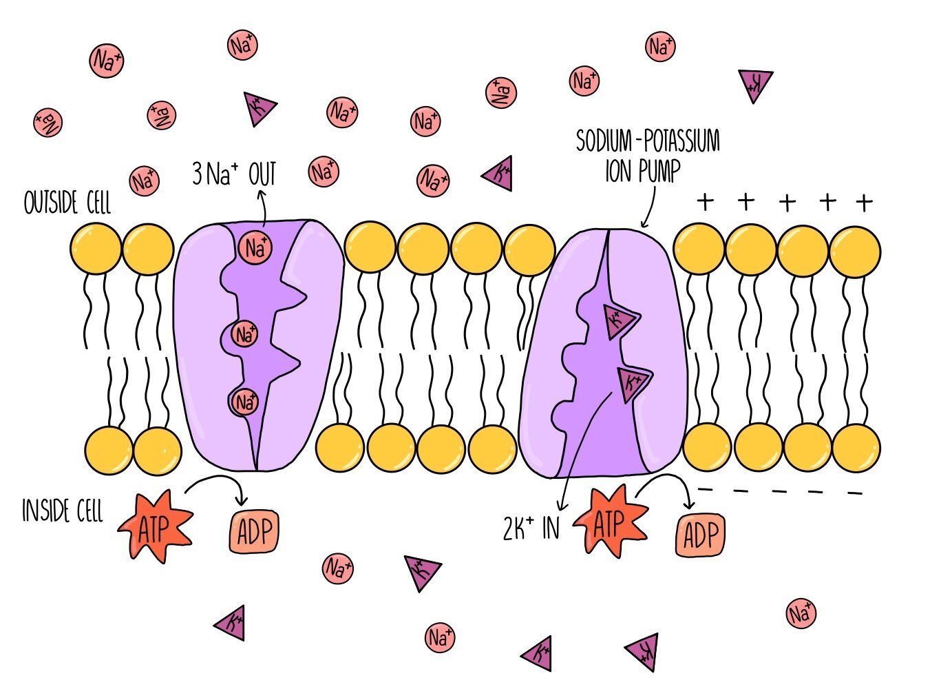
When a neurone is not firing (i.e. it is not transmitting an action potential), there is a difference in charge between the inside and the outside of the membrane (it is polarised). This charge difference is referred to as the resting potential and is usually around -70 mV. Polarisation of neuronal cell membranes at rest occurs due to the action of sodium-potassium ion pumps. These pumps are found within the cell membrane and actively transport sodium and potassium ions into and out of the neurone. For every three sodium ions that the proteins pump out of the cell, they pump two potassium ions into the cell. This ensures that there are always more positive ions out of the cell compared to inside the cell and makes sure there is a charge difference across the membrane.
Action potential
When a neurone is stimulated, the charge difference between the inside and outside of the cell membrane is lost and the membrane is depolarised. If enough charge is lost and depolarisation exceeds -55 mV, an action potential will occur in that neurone. The -55 mV ‘limit’ is known as the threshold potential - any depolarisation above this number will result in an action potential whereas anything less than that will result in nothing. We therefore refer to action potentials as an “all-or-nothing” response.
Depolarisation during an action potential occurs because sodium ion channels open up in the membrane. Remember that the sodium-potassium ion pump has been actively transporting sodium ions out of the neurone, creating a sodium ion concentration gradient. This means that when sodium ion channels open, sodium ions flood into the neuron by facilitated diffusion. The potential difference across the membrane is reduced until it reaches a voltage of around +30 mV.
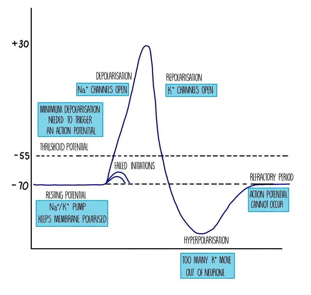
Sodium ion channels close and potassium ion channels open, which causes potassium ions to move out of the neurone down their concentration gradient. The movement of positive ions out of the cell means that there is a charge difference again across the membrane - this is called repolarisation. However, the charge difference exceeds the resting potential and becomes ‘hyperpolarised’. This is because the potassium ion channels are slow to close and too many potassium ions diffuse out of the neurone. The action of the sodium-potassium ion pump restores the balance between sodium and potassium ions on either side of the membrane and returns the neurone to its resting potential of -70 mV.
Immediately after an action potential is a brief period called the refractory period. During the refractory period, the neurone cannot be stimulated and an action potential cannot occur. This is because the ion channels are recovering and they cannot be made to open. The refractory period is important because it ensures that action potentials do not overlap (i.e. they pass along the neurone as separate impulses) and that action potentials are unidirectional.
Once an action potential occurs in one part of the neuron, it will stimulate an action potential in the adjacent part of the neuron, creating a kind of ‘Mexican wave’ of depolarisation. This wave of depolarisation occurs because the sodium ions which diffuse into the neuron diffuse sideways, causing voltage-gated ion channels in the next portion of the neurone to open, so sodium ions move into the neurone further along the membrane. The wave moves away from the part of the neurone which has just fired an action potential because that part of the neurone will be in the refractory period and cannot be stimulated.
Size of the stimulus
We’ve seen how an action potential is an ‘all-or-nothing’ response. If the threshold potential is reached, an action potential will occur. This action potential is always of the same voltage (depolarisation to +30 mV) regardless of whether the stimulus that initiated the action potential is small (e.g. a pinprick) or large (e.g. a sledgehammer). If the threshold isn’t reached then an action potential will not be fired. The difference between action potentials resulting from stimuli of different sizes is the frequency that action potentials are firing - the bigger the stimulus, the more often an action potential will occur along the neurone.
Factors that affect speed of an action potential
Myelination
Some neurones are insulated with a fatty layer along the axon. This fatty layer is called a myelin sheath and is made up of a type of cell called a Schwann cell.
The myelin sheath acts as an electrical insulator, which means that ions cannot move into or out of the myelinated portions of the neurone.
However, there are gaps in the myelin sheath called nodes of Ranvier, where sodium ion channels and potassium ion channels are concentrated.
Action potentials occur only at the nodes of Ranvier - when one node is stimulated this triggers depolarisation of the next node, causing the impulse to ‘jump’ from node to node.
This type of nervous transmission is called saltatory conduction and is much faster than transmission along non-myelinated neurones, where the action potential has to travel along the entire length of the neurone in a wave of depolarisation.
The speed at which an action potential moves along a neurone is known as the conduction velocity - the higher the conduction velocity, the faster the action potential is travelling. This means that action potentials along myelinated neurones have a higher conduction velocity compared to those travelling along non-myelinated neurones.
Axon diameter
Action potentials travel quicker in neurons with bigger axons because there is less resistance to the flow of ions than in a neuron with a thinner axon.
Less resistance means that depolarisation travels to the next portion of the axon sooner.
Temperature
As temperature increases, speed of an action potential increases because the ions have more kinetic energy so diffuse faster.
But this is only true up to a point – at temperatures above 40 degrees, conduction velocity slows due to the denaturation of channel and carrier proteins in the neuronal membrane.
Synapses
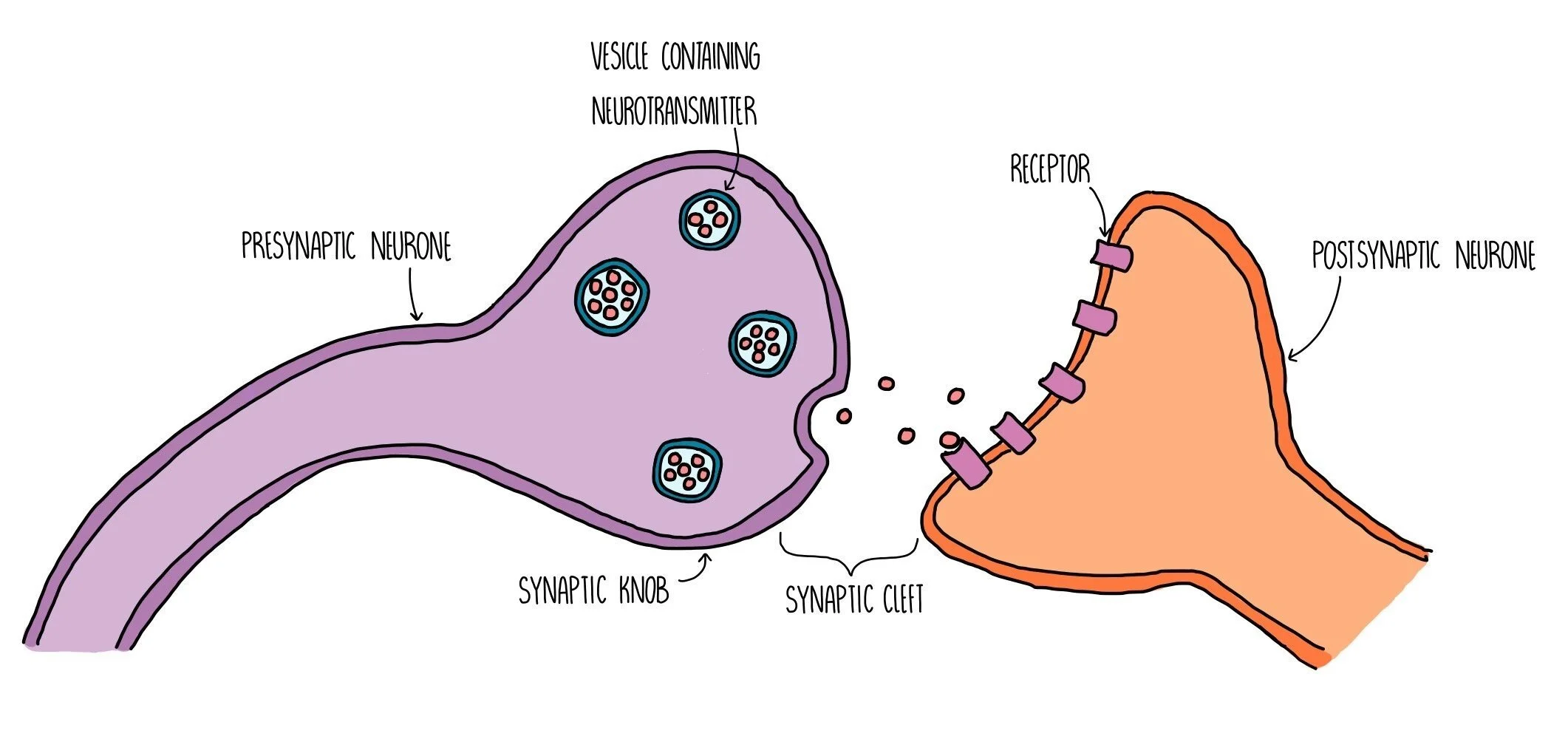
A synapse is a gap found between neurones (or between a motor neurone and an effector). Electrical impulses cannot pass through the gap, so neurones release neurotransmitters from one neurone to the next to stimulate an action potential in the next neurone. The neurone before the synapse is called the presynaptic neurone and the one after the synapse is called the postsynaptic neurone. The space between them is called the synaptic cleft. This means that action potentials will travel along the presynaptic neurone, through the synaptic cleft (via neurotransmitters) then along the postsynaptic neurone. The presynaptic neurone has a swelling at the end which is called the synaptic knob.
Synaptic transmission takes place in the following stages:
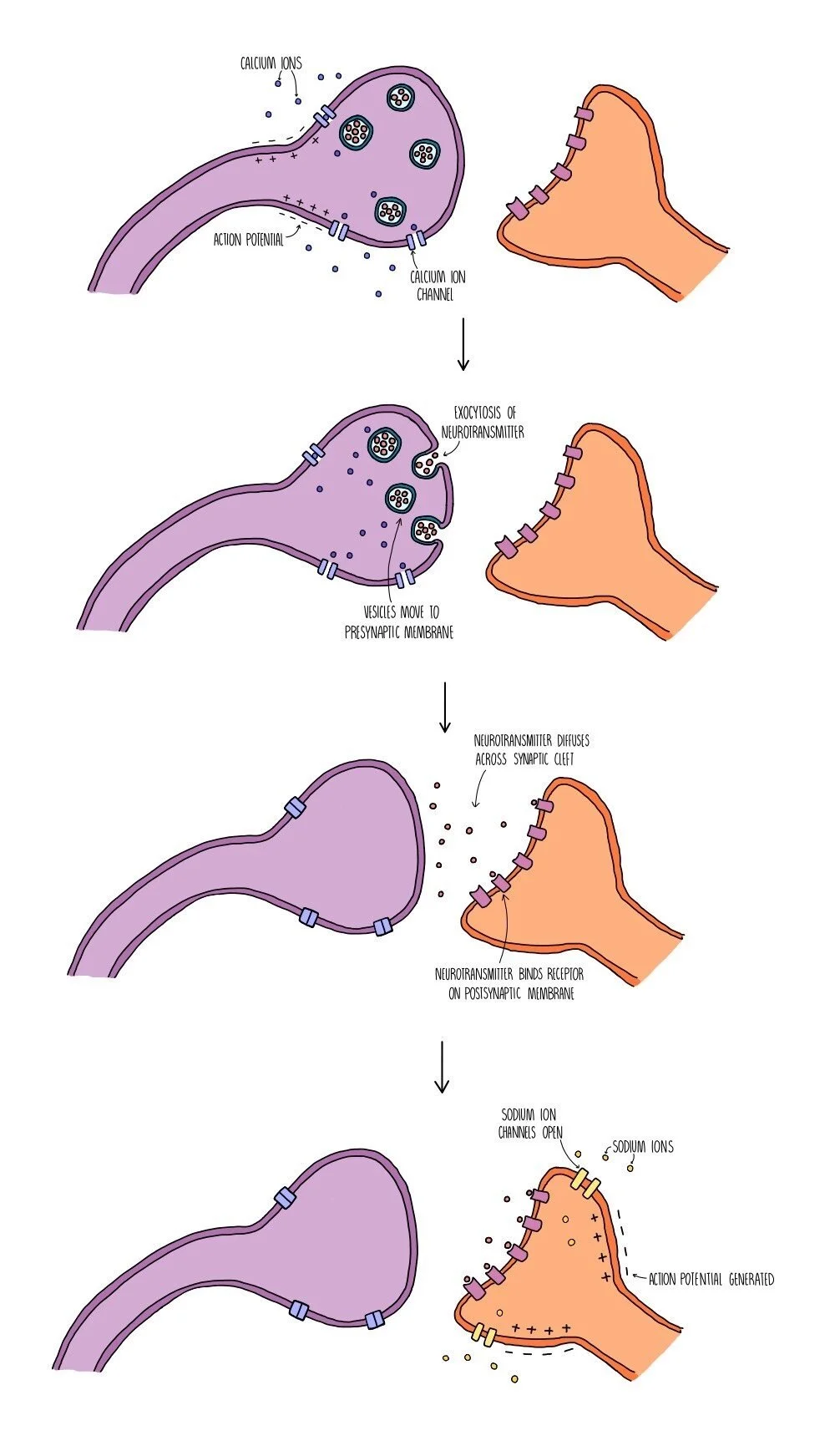
An action potential arrives at the end of the presynaptic neurone (at the synaptic knob) and triggers the opening of voltage-gated calcium ion channels.
Calcium ions move into the synaptic knob by facilitated diffusion and trigger the movement of vesicles containing neurotransmitters (such as acetylcholine or dopamine) towards the presynaptic membrane.
The vesicles fuse with the presynaptic membrane and their contents is released by exocytosis.
The neurotransmitters diffuse across the synaptic cleft and bind to specific receptors on the postsynaptic membrane.
This triggers the opening of sodium ion channels in the postsynaptic membrane. Sodium ions move into the postsynaptic neurone, causing depolarisation and triggering an action potential if the excitation exceeds the threshold potential of -55 mV.
The neurotransmitter is removed from the synaptic cleft which prevents the continuous stimulation of an action potential in the postsynaptic neurone. The neurotransmitter is either reabsorbed by the presynaptic neurone (and recycled) or broken down by enzymes in the synaptic cleft (and the products are reabsorbed).
Receptors which are complementary to neurotransmitters, such as acetylcholine, are only found on the postsynaptic membrane - there are none within the postsynaptic membrane). This ensures that the action potential always travels in one direction only (unidirectional) and prevents the nerve impulse from travelling backwards.
Cholinergic synapses
Synapses that use the neurotransmitter acetylcholine are called cholinergic synapses. They work in the same way as a typical synapse described above. Acetylcholine is broken down by the enzyme acetylcholinesterase and the products are reabsorbed into the presynaptic neuron to resynthesize the neurotransmitter.
Excitatory and inhibitory neurotransmitters
Neurotransmitters can be classed as excitatory if they trigger an action potential in the postsynaptic neuron or inhibitory if they prevent an action potential from happening. Some neurotransmitters are both excitatory and inhibitory, with their effect determined by where in the body they are acting. For example, acetylcholine is an excitatory neurotransmitter at cholinergic synapses in the CNS and neuromuscular junction. But in cholinergic synapses in the heart, acetylcholine acts as an inhibitory neurotransmitter by triggering the opening of potassium ion channels and making the membrane even more polarised (hyperpolarisation).
Summation
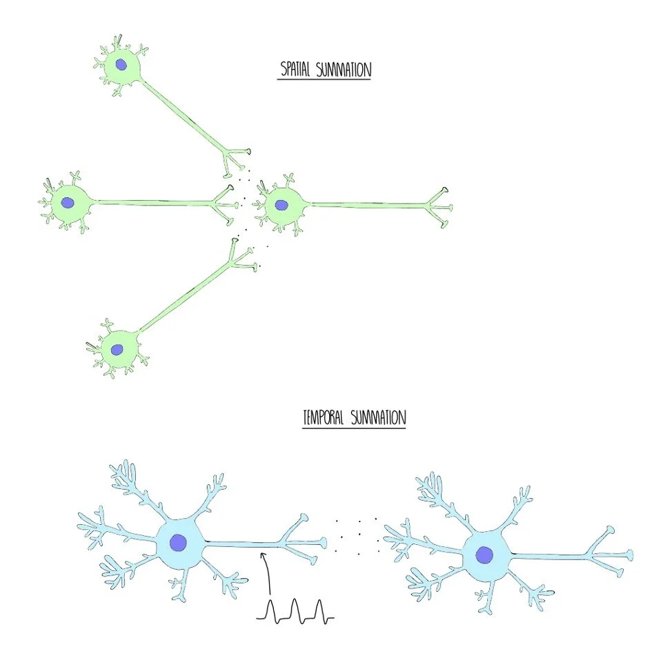
Summation is when small amounts of neurotransmitter build up to trigger an action potential in the postsynaptic neuron. Spatial summation occurs when lots of presynaptic neurons converge on a single postsynaptic neuron. Although each of the presynaptic neurons are releasing small amounts of neurotransmitter (which alone would not be enough to exceed the threshold potential), the combined amount is enough to stimulate an impulse in the next neuron.
Temporal summation is when a single neuron fires action potentials in quick succession, repeatedly releasing neurotransmitter into the synaptic cleft. This causes the amount of neurotransmitter in the synaptic cleft to increase, making an action potential in the postsynaptic neuron more likely.
Neuromuscular junctions
Neuromuscular junctions (NMJs) are the fancy name for a synapse between a motor neuron and a muscle cell. NMJs use the neurotransmitter acetylcholine and contain receptors called nicotinic cholinergic receptors. Unlike typical synapses:
The postsynaptic membrane in NMJs is folded into clefts which store the enzyme acetylcholinesterase, which breaks down acetylcholine.
Acetylcholine always acts as an excitatory neurotransmitter.
The postsynaptic membrane contains a higher number of receptors.
The effect of drugs
Drugs can have a range of effects on our body by interfering with neuronal signalling at the synapse. They can work in various ways:
Some bind to receptors and trigger an action potential in the postsynaptic neuron – this is because they are a similar shape to natural neurotransmitters and activate its receptors.
Some block receptors and prevent neurotransmitters from activating it.
Some inhibit enzymes that break down the neurotransmitter – this means that the neurotransmitter remains in the synaptic cleft and can continue stimulating the postsynaptic neuron.
Some trigger or inhibit the release of neurotransmitter from the presynaptic neuron.